Friday 7 September 2018

Eclectic Aproach : Latest Physiotherapy Technique :

  BASIC OF AN ELECTIC APPROACH :-

                                        Eclectic approach is a method of language education that combines various approaches and methodologies to teach language depending on the aims of the lesson and the   abilities of the learners. Different teaching methods are borrowed and adapted to suit the requirement of the learners. It breaks the monotony of the class. In addition, It is a conceptual                         approach that does not merely include one paradigm or a set of assumptions.

         # INTRODUCTION :- 

                                 
                                  There is little empirical evidence as to whether an integravite and eclectic approach or a specific concept approach is the most effective when working with children. It appears                         to the authors that there are a number of common factor to all approaches and philosophies , even where their implementation and emphasais may differ.
                                  They are :
                              > assessment and planning is necessary at all stages 
                              > early treatment is essential
                              > team work is important
                              > the child must be motivated and involved
                              > parents should be involved and supported.

         # ELECTISUM :-

                                Eclecticism is a conceptual approach that does not hold rigidly to a single paradigm or set of assumptions, but instead draws upon multiple theories, styles, or ideas to gain                       complementary insights into a subject, or applies different theories in particular cases. However, this is often without conventions or rules dictating how or which theories were                       combined.

                                It can sometimes seem inelegant or lacking in simplicity, and eclectics are sometimes criticized for lack of consistency in their thinking. It is, however, common in many fields of                       study. For example, most psychologists accept certain aspects of behaviorism, but do not attempt to use the theory to explain all aspects of human behavior.

                                Eclecticism in ethics, philosophy and religion is also known as syncretism.

          # APPROACH METHOD :-

                                      -  There are varied approaches and methods used for language teaching. In eclectic approach, the teacher can choose from these different methods and approaches:

                      > Grammar-translation Method: It is a method of teaching languages by which students learn grammatical rules and then apply those rules by translating between the target language                                                                             and the native language.

                      > Direct Method: In this method the teacher refrains from using the students' native language. The target language is directly used for teaching all the four skills—listening, speaking,                                                     reading and writing.

                      > Structural-situational Approach: In this approach, the teacher teaches language through a careful selection, gradation and presentation of vocabulary items and structures through                                                                                 situation based activities.

                      >  Audio-lingual/Audio-visual Method: In this style of teaching students are taught through a system of reinforcement. Here new words and grammar are directly taught without using the                                                                                       students' native language. However, unlike direct method, audio-lingual method does not focus on vocabulary. Instead, the teacher focuses on                                                                                              grammar through drill and practice.

                      > Bilingual Method: The word 'bilingual' means the ability to speak two languages fluently. In bilingual method, the teacher teaches the language by giving mother tongue equivalents of                                                          the words or sentences.

                      > Communicative Language Teaching: This approach lays emphasis on oral method of teaching. It aims to develop communicative competence in students.

                      > Total-Physical Response: It is based on the theory that memory is enhanced through association with physical response.

                      > The Silent Way: In this method the teacher uses a combination of silence and gestures to focus students' attention.

         # ADVANTAGES :- 
Eclectic Approach
 

                             > The teacher has more flexibility.
                             > No aspect of language skill is ignored.
                             > There is variety in the classroom.
                             > Classroom atmosphere is dynamic.
                             > These types of programs not only negotiate teacher skill-development within an improved recognition of and respect for cross-cultural and multi-linguistic classroom settings, but                                 also encourages student pride in their heritage, language, communication preferences and self-identity.

         # DISADVANTAGES :-
                             > It does not lend itself to prediction and control of behaviour.
                             > It's difficult to identify the relative contributions of each approach.
                             > Explanation of behaviour may become "watered down" when combining many perspectives.
                             > There are practical difficulties in investigating the integration of the approaches.
                             > It does not lend itself to hypothesis testing.
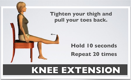
                            
         # DURING TREATMENT :-

Physiotherapy Treatment Technique
 

                              1. Neurodevlopmental sequences should be considered but not followed rigidly.
                              2. Postural mechanisms and normal postural tone should br developed involving
                                                    > postural fixation
                                                    > antigravity mechanisms
                                                    > righting reaction
                                                    >  equilibrium reactions.
                              3. Deformity should be prevented.
                              4. Afferent stimuli can be used
                                                    > touch
                                                    > temprature
                                                    > vision
                                                    > pressure
                                                    > stretch
                                                    > hearing
                              5. Sensorimotor experiences should be encouraged
                                                    > voluntary skilled movement
                                                    > cognition
                                                    > perception
                                                    > function
                              6. Gross motor activities generally precede fine motor movements.
                              7. Repetition & reinforcement are necessary
                              8. The devlopment of movement should lead to purposeful activity and independent function.

Saturday 14 July 2018

Physiotherapy Exercise Basic Rules :


Physiotherapy Exercise Some Basic Rules :

Basic Rules of Exercise :

Exercise is Require For Regular Interval is Require to Increase Stamina And Strength , But This Means Not to do anything , Exercise Are According to our

Body And Stamina And Under Proper Trainer or Physiotherapist Rules.
Here we Explain Basic Rules What to Care And Do's And Not To Do During , before And after Exercise.
Here we mentioned Some Simple 10 Rules To Take Notice During Exercise.

1.Proper Clothing :

  Proper Stretchable Clothing is Require During Exercise And Sports shoes During Brisk walking or Running.
  Do not wear any instrument like wrist watch, Ring And Related Any.

2. Do Exercise in Free Air And Open Space Or Room Have Proper Ventilated Because During Exercise Oxygen Require More.

3. Do Warm Up And Simple Stretching Before Exercise To Avoid Injury.

4. Do Not Do Too Much Exercise On Early Days, Gradually Increase Exercise.Do Take Enough Rest In Between Exercise.

5. Do Exercise in Early Morning And Evening is Good Time For Exercise.

6. To Avoid Dehydration Take Short Rest And Drink Some Water.

7. Don's do anything Over Exercise May Lead To Joint Pain Or Muscle Pain Or Related Sprain or Strain.

8. Follow Your Trainer Instruction Properly.

9. Avoid Exercise When You Feel Fatigue or Tired Or Not Something Well.

10. Do Proper Concentration , Outside Attention May Lead to Down Your Intensity Of Exercise And Sometimes Injury Too.


Exercise Therapy

Thursday 19 April 2018

MYOSITIS OSSIFICANS & PHYSIOTHERAPY :

DESCRIPTION :
X-Ray Shows Myositis Ossification in Thigh Area

Patient Of Myositis Osiification in Lower Limb


Myositis ossificans (MO) occurs when bone or bone-like tissue grows where it’s not supposed to. It most commonly happens in your muscle after an injury — like when you get hit hard in the thigh during a soccer game or maybe after a car or bicycle accident.Myositis ossificans comprises two syndromes characterized by heterotopic ossification (calcification) of muscle.About 80 percent of the time, these bony growths develop in the muscles of your thigh or upper arm.

CLASSIFICATION

In the first, and by far most common type, nonhereditary myositis ossificans, calcifications occur at the site of injured muscle, most commonly in the arms or in the quadriceps of the thighs.
The term myositis ossificans traumatica is sometimes used when the condition is due to trauma. Also known as Myositis ossificans circumscripta is another synonym of myositis ossificans traumatica refers to the new extraosseous bone that appears after trauma.
The second condition, myositis ossificans progressiva (also referred to as fibrodysplasia ossificans progressiva) is an inherited affliction, autosomal dominant pattern, in which the ossification can occur without injury, and typically grows in a predictable pattern. Although this disorder can be passed to offspring by those afflicted with FOP, it is also classified as nonhereditary, as it is most often attributed to a spontaneous genetic mutation upon conception.

CAUSES

Myositis ossificans usually occurs where a person has experienced a single traumatic injury, such as sustaining a hit while playing football or soccer that causes a deep muscle bruise.

It can also happen when there is a repetitive injury to the same area, such as in the thighs of horseback riders.

Sports injuries or accidents usually initiate MO. Adolescents and young adults in their 20s are most likely to develop MO. It’s rare for children age of 10 and under to get the condition. People who have paraplegia are also prone to develope MO, but usually with no evidence of trauma.

SIGNS & SYMPTOMS :


Symtom's Of Myositis Ossification


Unlike other typical muscle strains or injuries, people with myositis ossificans may notice that their pain worsens with time instead of getting better.
Someone with this condition may also notice changes in the affected muscle, including:

  • Warmth
  • Swelling
  • A lump or bump
  • Decreased range of motion
  • Tenderness

DIAGNOSIS :

X-Ray Myositis Ossification


If it has been at least 2–3 weeks since the pain or other symptoms started, the doctor may order some imaging tests to look for evidence of bone growth in the soft tissue.
Your doctor may also order other imaging tests. These may include a diagnostic ultrasound, MRI, CT, or bone scan.

X-ray: It can be difficult to diagnose myositis ossificans in the early stages with just an X-ray. Most X-rays will not show up any abnormalities in the first 2–3 weeks following the injury but will show changes after 3–4 weeks.

Ultrasound: Ultrasounds use sound waves to look at the soft tissues. They are one early diagnostic test that can be used to look for the changes associated with myositis ossificans. Ultrasonography depends on the ability of the person reading the scans, so many doctors do not often recommend it as the first test.

CT scan: Doctors can usually see the early development of bone tissue in soft tissues. However, it is not 100 percent reliable, and if a doctor suspects that someone has myositis ossificans, they may carry out additional testing to make the diagnosis.

Magnetic resonance imaging (MRI): An MRI is a preferred method of looking at soft tissue growths. A doctor may still order additional tests to compare and confirm a diagnosis.

A biopsy of the growth may also be taken and evaluated in a lab.


MANAGEMENT

  • Rest
  • Immobilization
  • Anti-inflammatory drugs
  • physiotherapy management
  • surgical debridement

Myositis ossificans usually resolves on its own.You may be able to prevent MO by properly taking care of your injury in the first two weeks. You can reduce inflammation by immobilizing the affected muscle with slight compression, icing, and elevation.

Rest: You don’t have to just lie there, but don’t stress the muscle too much.
Ice: Apply for 15 to 20 minutes at a time.
Compression: Wrap an elastic bandage firmly around your injury to minimize swelling and keep the area stable.
Elevation: Raise your injured limb above the level of your heart to help drain excess fluid from the area.
Non-painful stretching and strengthening: Gently stretch the affected muscle and start doing strengthening exercises when your doctor says it’s OK. Don’t perform any movements to the point of pain.

Medications and orthotics :

Physiotherapy Treatment


You can take nonsteroidal anti-inflammatory drugs like ibuprofen (Advil) or naproxen (Aleve) to reduce pain and swelling. Topical treatments like Biofreeze or Tiger Balm can also helps to ease pain.

When your pain and movement allow you to get back to sports, wear some padding or other protection on the injured muscle to prevent additional damage.

Physiotherapy management of myositis ossificans includes

Rest

Icing the injury

Pulsed Ultra sound and phonophoresis :

Electrotherapy in MO


Maintain available range of motion but avoid stretching and massage, until maturation.
Passive range of motion and mobilization: This is when a person or machine moves your body parts for you.
Active range of motion and mobilization: This is when you use your own strength to move your body parts.

Iontophoresis with 2 % acetic acid solution.

Extra corporeal shock wave therapy

Surgical Management

Growth should not be removed in premature stage as it will likely reoccur. The ossification becomes exuberant, infiltrates beyond the original site, and compresses the soft tissues around beyond hope of repair. When after serial x-rays the mass is dense, well delineated, and at a stand still, it may be safely removed. It may be possible to prevent myositis by aspirating the original haematoma.

PREVENTION :


Exercise in Myositis Ossification


While it can be difficult to predict who will get myositis ossificans, it is important to treat every injury promptly using the R.I.C.E. method. This is:

  • Rest
  • Ice
  • Compression
  • Elevation

An athlete who sustains an injury may need to leave the game or event, especially if there is significant swelling or bruising.


Ice Pack Treatment


Gentle stretching and range of motion exercises are also essential after an injury; myositis ossificans is more likely to affect a muscle that is not being used.

Doing too much too soon can worsen MO. But not working to recover your range of motion when the doctor says it’s safe may make your pain and stiffness last longer.

Monday 26 March 2018

Frankele's Co-ordination Exercise in Cerebeller Ataxia :

FRENKLE 'S  CO-ORDINATION    EXERCISE   FOR  CEREBELLER  ATAXIA :


It is the ability to execute smooth, accurate, controlled motor responses (optimal interaction of muscle function).

Coordination is the ability to select the right muscle at the right time with proper intensity to achieve proper action.
Coordinated movement is characterized by appropriate speed, distance, direction, timing and muscular tension.
It is the process that results in activation of motor units of multiple muscles with simultaneous inhibition of all other muscles in order to carry out a desired activity

Importance of the cerebellum in coordination ;

The cerebellum is the primary center in the brain for coordination of movement.

Components of coordinated movement:

Volition: is the ability to initiate,maintain or stop an activity or motion.
Perception:in tact proprioception and subcortical centres to integrate motor impulses and the sensory feedback. When proprioception is affected it is compensated with visual feedback.
Engramformation:is the neurologica lmuscular activity developed in the extrapyramidal system. Research proved that high repetitions of precise performance must be performed in order to develop an engram
.
Types of c-oordination:

1) Fine motor skills:

Require coordinated movement of small muscles (hands, face).
Examples: include writing, drawing, buttoning a shirt, blowing bubbles

2) Gross motor skills:

Require coordinated movement of large muscles or groups of muscles (trunk, extremities).
Examples: include walking, running, lifting activities.

3)Hand-eye skills:

The ability of the visual system to coordinate visual information. Received and then control or direct the hands in the accomplishment of a task .
Examples : include catching a ball,sewing,computer mouse use.

Causes of coordination impairments , Causes of Ataxia

Degeneration, damage or loss of nerve cells in the cerebellum, which is that part of the brain that controls muscle coordination, causes ataxia. The cerebellum comprises of two small ball-shaped folded tissues present at the base of the brain near the brainstem. Diseases which damage the spinal cord and peripheral nerves which connect the cerebellum to the muscles can also cause ataxia
.
 Other causes of ataxia include:

Stroke is a condition where the blood supply to a part of the brain gets severely diminished or interrupted, which deprives the brain tissue of oxygen and other nutrients resulting in death of brain cells.

Trauma or injury to the head, which causes damage to the brain or spinal cord, can cause sudden-onset ataxia (acute cerebellar ataxia).

Chickenpox can result in a complication in the form of Ataxia; although this is not common. Ataxia can appear during the healing stages of the infection and persist for days to weeks and gradually resolve over the time.

Transient ischemic attack (TIA) is caused by a temporary reduction in blood supply to a part of the brain. Majority of the TIAs last only for a few minutes. Some of the symptoms of TIA include ataxia, which is temporary.

Multiple sclerosis is a chronic, potentially debilitating medical condition, which affects the central nervous system.

Cerebral palsy consists of a group of disorders, which occurs as a result of damage to a child's brain during its early development. It can be before, during or shortly after birth. It affects the ability to coordinate movements of the body.

Paraneoplastic syndromes are rare, degenerative disorders, which are triggered by the response of the immune system to a tumor or neoplasm. This tumor is commonly in the lungs, ovaries, lymph nodes or breast. Patient can experience ataxia many months or years before cancer is actually diagnosed.

Toxic reaction to some medications can also cause ataxia. Medicines, especially barbiturates and certain sedatives, like benzodiazepine, can cause ataxia as a side effect. Other things, which could cause toxic reactions, are heavy metal poisoning, alcohol and drug intoxication and solvent poisoning.

Any type of growth on the brain, either cancerous or noncancerous, can damage cerebellum and cause ataxia.

Deficiency of vitamin E or B-12 can also lead to ataxia.

No specific cause can be found for some adults who develop sporadic ataxia, also known as sporadic degenerative ataxia, which can be of many types, such as multiple system atrophy which is progressive and degenerative disorder.

Examples of coordination tests:


1) In the upper limb:
Finger To Nose Test in Upper Limb


A) Finger-to-nose test

The shoulder is abducted to 90o with the elbow extended, the patient is asked to bring the tip of the index finger to the tip of the nose. Finger to therapist's finger: the patient and the therapist site opposite to each other, the therapist index finger is held in front of the patient, the patient is asked to touch the tip of the index finger to the therapist index finger.

B) Finger-to-finger test

Both shoulders are abducted to bring both the elbow extended, the patient is asked to bring both the hand toward the midline and approximate the index finger from the opposing hand

C) Finger-to-doctor's finger test

the patient alternately touches the tip of the nose and the tip of the therapist's finger with the index finger.

D) Adiadokokinesia or dysdiadokokinesia:

The patient was asked to do rapidly alternating movements e.g. forearm supination and pronation, hand tapping.

E) Rebound phenomena:

The patient with his elbow fixed, flex it against resistance. When the resistance is suddenly released the patient's forearm flies upward and may hit his face or shoulder.

F) Buttoning and unbuttoning test.

In any of the previous tests, we may find:

Intention tremors and Decomposition of movements
Dysmetria: in the form of hypermetria or hypometria

2) In the lower limb :

Heel To Sheen Test in Lower Limb


A) Heel-to-knee test

B) Walking along a straight line. Foot close to foot:In case of cerebellar lesion, there is deviation of gait

C) Rom-berg test: Ask the patient to stand with heels together. Swaying or loss of balance occurs while his eyes are open or closed.

General principles of coordination exercises involve:

Constant repetition of a few motor activities
Use of sensory cues (tactile, visual,proprioceptive) to enhance motor performance
Increase of speed of the activity over time
Activities are broken down into components that are simple enough to be performed correctly.
Assistance is provided when ever necessary.
The patient there fore should have a short rest after two or three repetitions,to avoid fatigue.
High repetition of precise performance must be performed for the engram to form.
When ever a new movement is trained, various inputs are given, like instruction(auditory), sensory stimulation(touch) ,or positions in which the patient can view the movement (visual stimulation) to enhance motor performance.

Therapeutic exercises used to improve coordination:

Frenkel’s exercises
Proprioceptive Neuromuscular Facilitation
Neurophysiological Basis of Developmental techniques
Sensory Integrative Therapy

FRENKEL’S EXERCISES:   

Frenkel aimed at establishing voluntary control of movement by the use of any part of the sensory mechanism which remained intact, notably sight, sound and touch, to compensate for the loss of kinaesthetic sensation.

The process of learning this alternative method of control is similar to that required to learn any new exercise,

the essentials being: Concentration of the attention, Precision and Repetition

The ultimate aim is to establish control of movement so that the patient is able and confident in his ability to carry out these activities which are essential for independence in everyday life.


They are a system of slow repetitious exercises. They increase in difficulty over the time of the program. The patient watches his hand or arm movements (for example) and corrects them as needed.

Although the technique is simple, needs virtually no exercise equipment, and can be done on one's own, concentration and some degree of perseverance is required. Research has shown that 20,000 to 30,000 repetitions may be required to produce results. A simple calculation will show that this can be achieved by doing 60 repetitions every hour for six weeks in a 16-hour daily waking period. The repetitions will take just a few minutes every hour.

The brain as a whole learns to compensate for motor deficits in the cerebellum (or the spinal cord where applicable). If the ataxia affects say, head movements, the patient can use a mirror or combination of mirrors to watch their own head movements.

History

Best Physiotherapy Exercises for In-Coordination--Frenkel’s Exercises :

Co-Ordination Exercise


Frenkel Exercises are a series of motions of increasing difficulty performed by ataxic patients to facilitate the restoration of coordination. Frenkel's exercises are used to bring back the rhythmic, smooth and coordinated movements.


Dr. H S Frenkel was a physician from Switzerland who aimed at establishing voluntary control of movement by the use of any part of the sensory mechanism which remained intact, notably sight, sound and touch, to compensate for the loss of kinaesthetic sensation.

Frenkel Exercises were originally developed in 1889 to treat patients of tabes dorsalis and problems of sensory ataxia owing to loss of proprioception. These exercises have been applied in the treatment of individuals with ataxia, in particular cerebellar ataxia. The exercises are performed in supine, sitting, standing and walking. Each activity is performed slowly with the patient using vision to carefully guide correct movement. These exercises require a high degree of mental concentration and effort. For those patients with the prerequisite abilities, they may be helpful in regaining control of movement through cognitive compensation strategies. Patients with partial sensation can progress to practicing exercises with eyes closed. The main principles of Frenkel exercises are the following:

    Concentration or attention
    Precision
    Repetition

This program consists of a planned series of exercises designed to help patient compensate for the inability to tell where the arms and legs are- in space without looking.

    1. Exercises are designed primarily for coordination; they are not intended for strengthening.
    2. Commands should be given in an event, slow voice; the exercises should be done to count.
    3. It is important that the area is well lit and that patients are positioned so that they can watch the movement of their legs.
    4. Avoid fatigue. Perform each exercise not more than four times. Rest between each exercise.
    5. Exercises should be done within a normal range of motion to avoid over-stretching of muscles.
    6. These simple exercises should be adequately performed before progressing to more difficult patterns.

General Instructions for frenkel exercises

    Exercises can be performed with the part supported or unsupported, unilaterally or bilaterally.
    They should be practiced as smooth, timed movements, performed at a slow, even tempo by counting out loud.
    Consistency of performance is stressed and a specified target can be used to determine range.
    Four basic positions are used: lying, sitting, standing and walking.
    The exercises progress from postures of greatest stability (lying, sitting) to postures of greatest challenge (standing, walking).
    As voluntary control improves, the exercises progress to stopping and starting on command, increasing the range and performing the same exercises with eyes closed.
    Concentration and repetition are the keys to success.

Frenkel exercises for lower limb

Exercises for the legs in lying

    Flex and extend one leg by the heel sliding down a straight line on the table.
    Abduct and adduct hip smoothly with knee bent and heel on the table.
    Abduct and adduct leg with knee and hip extended by sliding the whole leg on the table.
    Flex and extend hip and knee with heel off the table.
    Flex and extend both the legs together with the heel sliding on the table.
    Flex one leg while extending the other.
    Flex and extend one leg while taking the other leg into abduction and adduction.
    Heel of one limb to opposite leg (toes, ankle, shin, patella).
    Heel of one limb to opposite knee, sliding down crest of tibia to ankle.

Whether the patient slides the heels or lifts it off the bed he has to touch it to the marks indicated by the patient on the plinth. The patient may also be told to place the heel of one leg on various points of the opposite leg under the guidance of the therapist.
Exercises for the legs in Sitting

    One leg is stretched to slide the heel to a position indicated by a mark on the floor.
    The alternate leg is lifted to place the heel on the marked point.
    From stride sitting posture patient is asked to stand and then sit.
    Rise and sit with knees together.
    Sitting hip abduction and adduction.

Exercises for the legs in Standing

    In stride standing weight is transferred from one foot to other.
    Place foot forward and backward on a straight line.
    Walk along a winding strip.
    Walk between two parallel lines
    Walk sideways by placing feet on the marked point.
    Walk and turn around
    Walk and change direction to avoid obstacles.


Frenkel exercises for upper limb :

Similar exercises can be devised for the upper limb wherein the patient may be directed to place the hand on the various points marked on the table or wall board to improve coordination of all the movements in the upper limb.
Some examples of Frankel exercises of upper limb in sitting position

    Have patient sit in front of a table and place a number of objects on the table. The patient then touches each object with the right hand and then the left hand.
    The patient flexes the right shoulder to 90 degree with elbow and wrist extended. The patient then takes his or her right index finger and touches the tip of his or her nose. This exercise is then repeated with the left hand. The exercise is performed alternating right and left index finger.
    The patient taps bilateral hands on bilateral thighs while alternating palmer and dorsal surfaces as fast as possible.

Certain diversional activities such as building with toy bricks or drawing on a black board, buttoning, combing, writing, typing are some of the activities that also improves the coordination.


Monday 5 March 2018

Knee Joint : Detail And Basic Overview

Knee Joint: It’s Important : 


Knee Joint
Knee Joint Detail


 The knee joint is one of the strongest and most important joints in the human body. It allows the lower leg to move relative to the thigh while supporting the body’s weight. Movements at the knee joint are essential to many everyday activities, including walking, running, sitting and standing.
The knee, also known as the tibiofemoral joint, is a synovial hinge joint formed between three bones: the femur, tibia, and patella. Two rounded, convex processes (known as condyles) on the distal end of the femur meet two rounded, concave condyles at the proximal end of the tibia.
The patella lies in front of the femur on the anterior surface of the knee with its smooth joint-forming processes on its posterior surface facing the femur.
The joint-forming surfaces of each bone are covered in a thin layer of hyaline cartilage that gives them an extremely smooth surface and protects the underlying bone from damage. Between the femur and tibia is a figure-eight-shaped layer of tough, rubbery fibrocartilage known as the meniscus. The meniscus acts as a shock absorber inside the knee to prevent the collision of the leg bones during strenuous activities such as running and jumping.

As with all synovial joints, a joint capsule surrounds the bones of the knee to provide strength and lubrication. The outer layer of the capsule is made of fibrous connective tissue continuous with the ligaments of the knee to hold the joint in place. Oily synovial fluid is produced by the synovial membrane that lines the joint capsule and fills the hollow space between the bones, lubricating the knee to reduce friction and wear.
Tendons connect the knee bones to the leg muscles that move the knee joint. Ligaments join the knee bones and provide stability to the knee:


The anterior cruciate ligament prevents the femur from sliding backward on the tibia (or the tibia sliding forward on the femur).
The posterior cruciate ligament prevents the femur from sliding forward on the tibia (or the tibia from sliding backward on the femur).
The medial and lateral collateral ligaments prevent the femur from sliding side to side.
Two C-shaped pieces of cartilage called the medial and lateral menisci act as shock absorbers between the femur and tibia.
Numerous bursae, or fluid-filled sacs, help the knee move smoothly


Anatomy of Knee Joint:-


Bones:-

The femur (thigh bone), tibia (shin bone), and patella (kneecap) make up the bones of the knee. The knee joint keeps these bones in place.

The patella is a small, triangle shaped bone that sits at the front of the knee, within the quadriceps muscle. It is lined with the thickest layer of cartilage in the body because it endures a great deal of force.


Knee joint Anatomy
knee joint anatomy


Cartilage:-

There are two types of cartilage in the knee:

Meniscus: these are crescent-shaped discs that act as a cushion, or “shock absorber” so that the bones of the knee can move through their range of motion without rubbing directly against each other. The menisci also contain nerves that help improve balance and stability and ensure the correct weight distribution between the femur and tibia.

The knee has two menisci:

medial – on the inner side of the knee, this is largest of the two
lateral – on the outer side of the knee
Articular cartilage: found on the femur, the top of the tibia, and the back of the patella; it is a thin, shiny layer of cartilage. It acts as a shock absorber and helps bones move smoothly over one another.

Ligaments:-

Knee Joint SideView


Ligaments are tough and fibrous tissues; they act like strong ropes to connect bones to other bones, preventing too much motion and promoting stability. The knee has four:

ACL (anterior cruciate ligament) – prevents the femur from sliding backward on the tibia, and the tibia from sliding forward on the femur.
PCL (posterior cruciate ligament) – prevents the femur from sliding forward on the tibia, or the tibia from sliding backward on the femur.
MCL (medial collateral ligament) – prevents side to side movement of the femur.
LCL (lateral collateral ligament) – prevents side to side movement of the femur.

Tendons:-

These tough bands of soft tissue provide stability to the joint. They are similar to ligaments, but instead of linking bone to bone, they connect bone to muscle. The largest tendon in the knee is the patellar tendon, which covers the kneecap, runs up the thigh, and attaches to the quadriceps.

Muscles:-

Although they are not technically part of the knee joint, the hamstrings and quadriceps are the muscles that strengthen the leg and help flex the knee.

The quadriceps are four muscles that straighten the knee. The hamstrings are three muscles at the back of the thigh that bend the knee.

The gluteal muscles – gluteus medius and minimus – also known as the glutes are in the buttocks; these are also important in positioning the knee.

Joint capsule:-

The joint capsule is a membrane bag that surrounds the knee joint. It is filled with a liquid called synovial fluid, which lubricates and nourishes the joint.

Bursa:-

There are approximately 14 of these small fluid-filled sacs within the knee joint. They reduce friction between the tissues of the knee and prevent inflammation.

* Knee Conditions:-

(1)Chondromalacia patella (also called patellofemoral syndrome): Irritation of the cartilage on the underside of the kneecap (patella), causing knee pain. This is a common cause of knee pain in young people.
(2)Knee osteoarthritis: Osteoarthritis is the most common form of arthritis, and often affects the knees. Caused by aging and wear and tear of cartilage, osteoarthritis symptoms may include knee pain, stiffness, and swelling.
(3)Knee effusion: Fluid buildup inside the knee, usually from inflammation. Any form of arthritis or injury may cause a knee effusion.
(4)Meniscal tear: Damage to a meniscus, the cartilage that cushions the knee, often occurs with twisting the knee. Large tears may cause the knee to lock.
(5)ACL (anterior cruciate ligament) strain or tear: The ACL is responsible for a large part of the knee’s stability. An ACL tear often leads to the knee “giving out,” and may require surgical repair.
(6)PCL (posterior cruciate ligament) strain or tear: PCL tears can cause pain, swelling, and knee instability. These injuries are less common than ACL tears, and physical therapy (rather than surgery) is usually the best option.
(7)MCL (medial collateral ligament) strain or tear: This injury may cause pain and possible instability to the inner side of the knee.
(8)Patellar subluxation: The kneecap slides abnormally or dislocates along the thigh bone during activity. Knee pain around the kneecap results.
(9)Patellar tendonitis: Inflammation of the tendon connecting the kneecap (patella) to the shin bone. This occurs mostly in athletes from repeated jumping.
(10)Knee bursitis: Pain, swelling, and warmth in any of the bursae of the knee. Bursitis often occurs from overuse or injury.
(11)Baker’s cyst: Collection of fluid in the back of the knee. Baker’s cysts usually develop from a persistent effusion as in conditions such as arthritis.
(12)Rheumatoid arthritis: An autoimmune condition that can cause arthritis in any joint, including the knees. If untreated, rheumatoid arthritis can cause permanent joint damage.
(13)Gout: A form of arthritis caused by a buildup of uric acid crystals in a joint. The knees may be affected, causing episodes of severe pain and swelling.
(14)Pseudogout: A form of arthritis similar to gout, caused by calcium pyrophosphate crystals depositing in the knee or other joints.
(15)Septic arthritis: An infection caused by bacteria, a virus, or fungus inside the knee can cause inflammation, pain, swelling, and difficulty moving the knee. Although uncommon, septic arthritis is a serious condition that usually gets worse quickly without treatment.

Prevention of knee injuries:-

Quadriceps Active Exercise
Stepping Exercise For Quadriceps Strenthening Exercise
Prone Knee Bending For Hamstring Active Exercise

The following tips may help prevent common knee injuries:

Warm-up by walking and stretching gently before and after playing sports.
Keep the leg muscles strong by using stairs, riding a stationary bicycle, or working out with weights.
Avoid sudden changes in the intensity of exercise.
Replace worn-out shoes. Choose ones that fit properly and provide good traction.
Maintain a healthy weight to avoid added pressure on the knees.
Always wear a seatbelt.
Use knee guards in sports where knees could get injured.
Maintaining strong, flexible leg muscles and seeking prompt medical attention for all knee injuries is essential to assure accurate diagnosis and appropriate treatment of the injury. Additionally, keeping the supporting leg muscles strong and practicing injury prevention will help keep the knee healthy across the lifespan.


Strengthening exercise for the knee:-


(1)Mini or partial squats with a chair or at a counter (quadriceps):

Holding on to a chair or stable surface, with knees about shoulder-width apart and pointing forward, slightly bend hips and knees as if sitting down onto a chair, and then slowly stand back up. Repeat 10 to 12 times.

(2)Standing hamstring curls (hamstrings):

Holding on to the back of a chair or stationary surface, without moving hip, bend knee as far as possible, bringing your heel up towards your buttocks. Do 10 to 12 reps on each leg.

(3)Marching in place (hip flexors and a good balance exercise):

On your own or while holding on to the back of a chair or stationary object, take alternating steps in place, bringing your knee up to a comfortable height. Strive for 60 seconds of marching.

(4)Heel raises (calf muscle):

Holding on to the back of a chair or stable surface, rise up on toes, lift heels off ground, and then slowly lower back down. Do 10 to 12 reps.

(5)Quad sets:

This simple exercise may be done on the floor with or without a pillow under your knee. Sit with your legs out in front of you and your knees completely straight (lean against a wall or back on your hands). Focus on contracting your quadriceps muscle and holding it as tight as possible for several seconds; relax and repeat 10 times. Repeat several times a day if your knees actively ache.

(6)Straight leg raises:

In the same starting position as the quad sets, sit with your right leg (do one at a time) straight in front of you with your toes pulled towards the knee. (If this is too difficult you may also do these lying on your back to start.) Keep your left leg bent with your foot on the floor. Contract your quads on your right leg, lift your foot about 12 inches off the ground and hold it up for 5 seconds; slowly lower it back down and repeat 10 times. Switch legs.

(7)Wall slides with ball squeeze:

Stand with your back against the wall and your feet shoulder-width apart. Hold a small (soccer ball size) inflated ball between your knees. Slowly slide down the wall by bending your knees and lowering yourself (knees should form a right angle with quads parallel to the floor and shins perpendicular to the floor). Hold 5 to 10 seconds and slowly return to starting position. Repeat 10 or more times.

(8)Clams:

Lie on your side with your hip and knee bent to approximately a 90-degree angle, with feet together. While keeping your ankles together, raise your top knee up about 12 inches from the other in a clamshell-type motion. Repeat 10 to 25 times and switch sides.

(9)Glute bridges:

Lie on your back with both knees bent at about a 90-degree angle with your feet on the floor. Tighten your buttocks as you lift your bottom off the floor as high as you can without arching your back; shoulders, hips, and knees should align. Hold this position as you extend one leg up while keeping knees aligned; hold 3 to 5 seconds and lower. Repeat on the opposite side. Perform 10 to 25 reps per side.

Include one or more of these exercises along with or instead of your usual leg routine two to three times a week for stronger legs and healthier, pain-free knees.

Famous surgery for knee joint:-


(1)Arthroscopy:-

In knee arthroscopy, a surgeon will look inside the knee joint, repair torn ligaments and remove damaged parts. Two or three very small incisions are made on the front of the knee. A fiber-optic camera is inserted through one incision. A surgical instrument is inserted through the other incision.

The surgeon can then examine and repair the knee. Knee scopes are most often performed for meniscal tears (torn cartilage). A degenerative tear can be debrided (cleaned up) during the arthroscopy. A traumatic sports-related tear can be debrided or repaired via arthroplasty.

Because of the minimal soft tissue damage resulting from an arthroscopy, recovery is relatively quick. It is a relatively easy surgery and most patients go home immediately after the scope. Patients will typically be able to resume normal activity and return to work within two or three weeks. The knee will be swollen for less than a week.

(2)Osteotomy:-

Knee osteotomy is a surgical procedure in which the surgeon removes or adds a wedge of bone to the top of your tibia (shinbone) or the bottom of your femur (thighbone). This provides a less worn area of articular cartilage to the weight-bearing part of the joint.

Osteotomy is typically recommended for those with arthritis damage in just one area of the knee. Arthritis on just one side of the knee can cause the knee to bow inward (valgus deformity) or outward (varus deformity). This can be corrected by the removal or addition of a wedge of bone. (Traumatic injury or even birth defects can also cause misalignment for which osteotomy is an appropriate surgical intervention.)

Many patients who undergo knee osteotomy will eventually need a total knee replacement. The osteotomy will buy them a varying amount of time before the need for total joint replacement becomes necessary.

Importance of knee joint:- 


why knee joint is so important in India?


We need our knees to run, walk, squat. With research suggesting our bones are weaker than those of Westerners, here’s a quiz to test how well you are caring for your ‘hinges’
Walking, running, climbing, dancing — the knees bear the brunt of every move we make throughout our lives. The main hinge between the ground and the body, knees bring together the femur (thigh bone), tibia (shin bone), fibula (next to tibia), and kneecap, and work as wheels that keep you going.

Sunday 18 February 2018

ORAL SUB MUCOUS FIBROSIS : Physiotherapy Treatment

ORAL SUB MUCOUS FIBROSIS :
A Patient with Oral submucosal Fibrosis


DEFINATION:

Oral submucous fibrosis is characterized as the unending, tricky ailment influencing the oral pit and here and there pharynx, albeit at times went before or potentially connected with vesicle arrangement and is constantly connected with juxtaepithelial fiery response took after by fibro versatile changes in the lamina propria with epithelial decay prompting firmness of oral pit prompting trismus and powerlessness to eat.

Oral submucous fibrosis is an interminable crippling and an all around perceived possibly threatening condition related with areca nut biting, an element of betel quid and is pervasive in South Asian populace. Pathogenesis isn't yet settled however is accepted to be because of multifactorial causes; consequently the treatment of oral submucous fibrosis proposes a noteworthy test for oral doctors.


ETIOLOGY AND PATHOPHYSIOLOGY:


The pathogenesis of the illness isn't entrenched, however, the reason for OSF is accepted to be multifactorial.

Various variables may trigger the infection procedure by causing a juxtaepithelial fiery response in the oral mucosa. Components incorporated are areca nut biting, ingestion of chilies, hereditary and immunologic procedures, wholesome inadequacies, and different variables.


Areca Nut (Betel Nut) Chewing:

The areca nut segment of betel quid assumes a noteworthy part in the pathogenesis of OSF 15. Betel nut is much of the time utilized as a psychotropic and antihelminthic operator and utilized as an after feast digestant which is taken to ease stomach inconvenience.

Smoking and liquor utilization alone, propensities basic to areca nut chewers, have been found to have no impact in the advancement of OSF. The most grounded confirm in regards to the etiology of OSF is with the propensity for areca nut biting.

Areca nut shape might be accessible in the following structure:

Supari + Tobacco

Supari + Pan+ Tobacco

Supari + Pan + Pan masala

Skillet Parag/Pan masala

Supari + Pan + Lime

Supari-Roasted/Raw Areca nut

Part of areca nut in pathogenesis of OSF:

Arecoline, a dynamic alkaloid found in betel nuts. Animates fibroblasts to build creation of collagen by 150%.

To lift the mRNA and protein articulation of cystatin C, a nonglycosylated fundamental protein reliably up-directed the assortment of fibrotic illnesses, in a measurement subordinate way in people with OSF.

Areca nuts have likewise been appeared to have a high copper substance, and biting areca nuts for 5-30 minutes altogether increments solvent copper levels in oral liquids. This expanded level of solvent copper underpins the speculation as a starting element in people with OSF.

Healthful Deficiencies:

Press insufficiency frailty, vitamin B complex inadequacy and lack of healthy sustenance are advancing variables that unsettle the repair of the excited oral mucosa, prompting damaged recuperating and resultant scarring.

The resultant atrophic oral mucosa is more defenseless to the impacts of chilies and betel nuts. Mucosal changes like those in vitamin B and iron insufficiency are found in oral sub mucosal fibrosis.

Chillies:

The part of chillies ingestion in the pathogenesis of OSF is disputable.

A touchiness response to chilies is accepted to add to OSF.

Hereditary and Immunologic Processes:

A hereditary segment is thought to be engaged with OSF Patients with expanded recurrence of HLA-A10, HLA-B7, and HLA-DR3.

Phases OF OSF:

Stage 1:

Stomatitis incorporates erythematous mucosa, vesicles, mucosal ulcers, melanotic mucosal pigmentation, and mucosal petechia.

Stage 2:

Fibrosis happens in cracked vesicles and ulcers when they mend, which is the sign of this stage.

Early sores show whitening of the oral mucosa.

More established sores incorporate vertical and round unmistakable sinewy groups in the buccal mucosa and around the mouth opening or lips, bringing about a mottled, marble like appearance of the mucosa in view of the vertical, thick, stringy groups running in a whitening mucosa. Particular discoveries incorporate the accompanying:

Decrease of the mouth opening (trismus).

Solid and little tongue.

Whitened and rough floor of the mouth.

Fibrotic and depigmented gingiva.

Rubbery delicate sense of taste with diminished portability.

Whitened and atrophic tonsils.

Contracted budlike uvula.

Sinking of the cheeks, not comparable with age or nutritious status.

Stage 3:

Screech of OSF are as per the following:

Leukoplakia is precancerous and is found in over 25% of people with OSF.

Discourse and hearing deficiencies may happen in view of inclusion of the tongue and the eustachian tubes.

Manifestations:

Xerostomia.

Intermittent ulceration.

Torment in the ear or deafness.

Nasal pitch of voice.

Confinement of the development of the delicate sense of taste.

Diminishing and hardening of the lips.

Pigmentation of the oral mucosa.

Dryness of the mouth and consuming sensation.

Diminished mouth opening and tongue bulge.

CAUSES : 

Mouth Opening Device in SMF


Immunological ailments.

Outrageous climatic conditions.

Delayed insufficiency to iron and vitamins in the eating regimen.

DIFFERENTIAL DIAGNOSIS:


Oral appearances of scleroderma

Oral appearances of Plummer Vinson disorder (Iron lack Anemia).

Examination:

Finish Hemogram

Toludine blue test

Biopsy :- Incisional biopsy

Immunofluorescent test:

a) Direct b) Indirect

Administration AND PREVENTION:

The treatment of patients with OSF relies upon the level of clinical contribution. On the off chance that the malady is identified at a beginning period, suspension of the propensity is adequate. Most patients with OSMF give moderateto-serious arranging. Direct to-serious arranging of OSF is irreversible. 

Medicinal treatment is symptomatic and gone for enhancing mouth developments.

Not to devour areca nut and other incessant aggravation, for example, hot and zesty sustenance including chiles.

Guidance green verdant vegetables.

Organization of Vit. A, B complex and high protein consume less calories.

Organization of Antoxid OD for 6 – two months.

Organization of Lycored OD for 6 two months.

Keeping up legitimate oral cleanliness.

Supplementing the eating regimen with nourishments rich in vitamins A, B complex, and C and iron.

Swearing off hot liquids like tea, espresso.

Swearing off liquor.

Utilizing a dental specialist to round off sharp teeth and concentrate third molars.

SURGICAL MANAGEMENT:

Surgical treatment is shown in patients with extreme conditions. These incorporate:-

Basic extraction of the stringy groups: Excision can bring about contracture of the tissue and intensification of the condition.

Split-thickness skin joining following respective temporalis myotomy or coronoidectomy: Trismus related with OSF might be because of changes in the temporalis ligament auxiliary to OSF; subsequently, skin unions may assuage.

Nasolabial folds and lingual pedicle folds: Surgery performed just in patients with OSF in whom the tongue isn't included.

PHYSIOTHERAPY MANAGEMENT: 


Mouth Opening Exercise In SMF


Muscle extending practices for the mouth might be useful to anticipate advance restrictions of mouth opening strong mouth opening has been attempted with mouth choke and non-cyclic surgical screw.

Diathermy: Microwave diathermy appears to be better than short wave, in light of the fact that specific warming of juxtaepitheliel connective tissue is conceivable it acts by physio fibrinolysis of groups.

Ultrasound: Ultra sound selectivity bring the temperature up in some all around aggregated territories. Ultrasound turns out to be an effective profound warming methodology.