Showing posts with label Deformity. Show all posts

Monday, 15 August 2022

Lumbar Lordosis

What is Lumbar Lordosis?

Lumbar Lordosis

Lumbar lordosis is the inward curvature of the lumbar spine. You may notice that some people have a small degree of lordosis (swayback) while others (especially those with very upright sitting postures) have severe lumbar spine curvature. 

It can be caused by a number of conditions such as habitual harmful postures, injury, and fractures, especially after childbirth. The lumbar spine plays a vital role in supporting your body’s weight, and keeping you stable and normal curves serve to distribute mechanical stress incurred as the body is at rest and during movement.

Lumbar lordosis is the inward curve of the lumbar spine. A small degree of lumbar spine curvature is normal while too much lumbar spine curvature is called lordosis (swayback).

The lumbar spine is a part of the trunk that supports the organs in your lower body while controlling muscle movements. In addition, it is used for flexibility and movement energy transmission. A small degree of lordosis is normal while too much lumbar spine curving is called lordosis (swayback).

Lumbar lordosis can be caused by genetic factors, obesity, and pregnancy. The symptoms of lumbar lordosis are related to the severity of the condition. Some people may have no symptoms at all, while others may experience pain in their lower back, numbness in their legs or feet, and difficulty walking.

Symptoms of Lumbar Lordosis

Lumbar lordosis is a condition where the spine curves inwards at the lower back. 

This condition can cause pain in the lower back and hamper mobility.

 Common symptoms of lumbar lordosis include:

  • Lower back pain and/or stiffness
  • It can cause pain and discomfort in the lower back, hips, buttocks, and legs.
  • Hip pain and/or stiffness
  • Tingling or numbness in the feet or toes
  • Pain while sitting for long periods of time
  • Tightness of the hamstrings
  • A feeling of instability or insecurity
  • Difficulty standing up straight
  • Pain during sitting or standing for long periods of time

The symptoms of lumbar lordosis are related to the severity of the condition. Some people may have no symptoms at all, while others may experience pain in their lower back, numbness in their legs or feet, and difficulty walking.

What Causes Lumbar Lordosis?

Lumbar lordosis is the natural inward curve of the spine. It is caused by a combination of genetics, age, and muscle imbalance.

It is a condition in which the back becomes arched or curved. This can happen due to a number of reasons.

 Some causes of lumbar lordosis are:

  • Poor posture
  • sitting for long periods of time
  • working at a desk all-day
  • obesity
  • Some medical conditions such as osteoporosis, scoliosis, and spondylolisthesis

Risk Factor:


The risk factors for lumbar lordosis include 

  • poor posture, 
  • being overweight or obese
  • having weak abdominal muscles
  • Abdominal surgery

Diagnosis Lumbar Lordosis

The condition can be diagnosed by a physical examination, X-rays, and MRI scans. It can also be diagnosed with a special test called the standing flexion test or the standing extension test.

Diagnosis of lumbar lordosis is done through the following steps:

Physical examination: A physical examination can be done to identify the symptoms of lumbar lordosis. The doctor will check for any abnormalities in muscle strength, posture, or gait. The doctor will also examine for any visible signs of injury to the back and spinal cord.

X-ray: An x-ray of the spine can help diagnose lumbar lordosis as well by identifying any structural abnormalities such as scoliosis or spondylolisthesis.

MRI: An MRI scan can also be used to diagnose

How is Lumbar Lordosis Treated?

There are several ways to treat lumbar lordosis.

Medical treatment is mostly symptomatic if you have Pain, the Doctor prescribes you Pain relieving Medicine mainly Non-steroidal anti-inflammatory drugs (NSAIDs) and If your bone becomes weak (osteoporosis) they also give you Calcium with Vitamin d3 combination medicine.

The first option is to use physical therapy to strengthen the muscles around the area. The physiotherapist prescribes you a few exercises and tips that give you a permanent solution and it is a natural way to recover without any side effects.

Exercise

Exercise for Lumbar Lordosis

Exercise is one of the best ways to treat lumbosacral pain. You can try walking, swimming, cycling, running, dancing, yoga, Pilates, weight training, etc. If you have lumbar pain, you should avoid activities that require you to sit or stand for long periods of time.

Stretching exercise

Stretching is a great way to relieve back pain caused by lumber lordosis. Stretching helps to loosen tight muscles and increase flexibility. There are many different types of stretching exercises that can be performed to relieve back pain. 

One example of a stretching exercise is performing a forward bend while holding onto something for balance. Another example would be to lie down on the floor and lift both legs off the ground. These two examples are just a couple of ways to stretch out the back muscles.

Strengthening Exercise:

Strengthening exercises of weak Abdominal muscles improves lumbar curvature and makes your spine more stable and normal.

Massage Therapy

Massage therapy helps relax muscles and reduce tension. It can help improve blood circulation and lymphatic drainage. It can also help increase flexibility and range of motion.

Heat Packs

Heat packs are a great way to relieve muscle tension and promote healing. Heat packs can be placed directly over the affected area to provide relief.

Ice Pack

Ice packs are a great way for patients to relieve back pain. Ice packs are applied directly to the painful area and left on for about 15 minutes. After the ice pack has been removed, the area should be massaged to stimulate blood flow.

Acupuncture

Acupuncture is a traditional Chinese medicine technique that uses fine needles to stimulate points along the meridians of the body. Stimulating these points can have a positive effect on reducing back pain.
Load more 

Chiropractic Care

Chiropractic care focuses on the alignment of the spine. By adjusting the spine, chiropractors can help eliminate nerve interference and restore proper function.

Surgery is the last option you should select if Medical and Physical therapy do not help.

Conclusion: A Guide to Treating Back Pain Caused by Lumber Lordosis

In order to treat lumber lordosis, the patient should perform regular exercises that strengthen their core muscles and help them maintain proper posture. Lordotic posture mostly recovers with the Help of Physiotherapy treatment and exercise, However, you should contact a nearby Physiotherapist or Physician for the Best treatment.

Sunday, 18 February 2018

ORAL SUB MUCOUS FIBROSIS : Physiotherapy Treatment

ORAL SUB MUCOUS FIBROSIS :
A Patient with Oral submucosal Fibrosis


DEFINATION:

Oral submucous fibrosis is characterized as the unending, tricky ailment influencing the oral pit and here and there pharynx, albeit at times went before or potentially connected with vesicle arrangement and is constantly connected with juxtaepithelial fiery response took after by fibro versatile changes in the lamina propria with epithelial decay prompting firmness of oral pit prompting trismus and powerlessness to eat.

Oral submucous fibrosis is an interminable crippling and an all around perceived possibly threatening condition related with areca nut biting, an element of betel quid and is pervasive in South Asian populace. Pathogenesis isn't yet settled however is accepted to be because of multifactorial causes; consequently the treatment of oral submucous fibrosis proposes a noteworthy test for oral doctors.


ETIOLOGY AND PATHOPHYSIOLOGY:


The pathogenesis of the illness isn't entrenched, however, the reason for OSF is accepted to be multifactorial.

Various variables may trigger the infection procedure by causing a juxtaepithelial fiery response in the oral mucosa. Components incorporated are areca nut biting, ingestion of chilies, hereditary and immunologic procedures, wholesome inadequacies, and different variables.


Areca Nut (Betel Nut) Chewing:

The areca nut segment of betel quid assumes a noteworthy part in the pathogenesis of OSF 15. Betel nut is much of the time utilized as a psychotropic and antihelminthic operator and utilized as an after feast digestant which is taken to ease stomach inconvenience.

Smoking and liquor utilization alone, propensities basic to areca nut chewers, have been found to have no impact in the advancement of OSF. The most grounded confirm in regards to the etiology of OSF is with the propensity for areca nut biting.

Areca nut shape might be accessible in the following structure:

Supari + Tobacco

Supari + Pan+ Tobacco

Supari + Pan + Pan masala

Skillet Parag/Pan masala

Supari + Pan + Lime

Supari-Roasted/Raw Areca nut

Part of areca nut in pathogenesis of OSF:

Arecoline, a dynamic alkaloid found in betel nuts. Animates fibroblasts to build creation of collagen by 150%.

To lift the mRNA and protein articulation of cystatin C, a nonglycosylated fundamental protein reliably up-directed the assortment of fibrotic illnesses, in a measurement subordinate way in people with OSF.

Areca nuts have likewise been appeared to have a high copper substance, and biting areca nuts for 5-30 minutes altogether increments solvent copper levels in oral liquids. This expanded level of solvent copper underpins the speculation as a starting element in people with OSF.

Healthful Deficiencies:

Press insufficiency frailty, vitamin B complex inadequacy and lack of healthy sustenance are advancing variables that unsettle the repair of the excited oral mucosa, prompting damaged recuperating and resultant scarring.

The resultant atrophic oral mucosa is more defenseless to the impacts of chilies and betel nuts. Mucosal changes like those in vitamin B and iron insufficiency are found in oral sub mucosal fibrosis.

Chillies:

The part of chillies ingestion in the pathogenesis of OSF is disputable.

A touchiness response to chilies is accepted to add to OSF.

Hereditary and Immunologic Processes:

A hereditary segment is thought to be engaged with OSF Patients with expanded recurrence of HLA-A10, HLA-B7, and HLA-DR3.

Phases OF OSF:

Stage 1:

Stomatitis incorporates erythematous mucosa, vesicles, mucosal ulcers, melanotic mucosal pigmentation, and mucosal petechia.

Stage 2:

Fibrosis happens in cracked vesicles and ulcers when they mend, which is the sign of this stage.

Early sores show whitening of the oral mucosa.

More established sores incorporate vertical and round unmistakable sinewy groups in the buccal mucosa and around the mouth opening or lips, bringing about a mottled, marble like appearance of the mucosa in view of the vertical, thick, stringy groups running in a whitening mucosa. Particular discoveries incorporate the accompanying:

Decrease of the mouth opening (trismus).

Solid and little tongue.

Whitened and rough floor of the mouth.

Fibrotic and depigmented gingiva.

Rubbery delicate sense of taste with diminished portability.

Whitened and atrophic tonsils.

Contracted budlike uvula.

Sinking of the cheeks, not comparable with age or nutritious status.

Stage 3:

Screech of OSF are as per the following:

Leukoplakia is precancerous and is found in over 25% of people with OSF.

Discourse and hearing deficiencies may happen in view of inclusion of the tongue and the eustachian tubes.

Manifestations:

Xerostomia.

Intermittent ulceration.

Torment in the ear or deafness.

Nasal pitch of voice.

Confinement of the development of the delicate sense of taste.

Diminishing and hardening of the lips.

Pigmentation of the oral mucosa.

Dryness of the mouth and consuming sensation.

Diminished mouth opening and tongue bulge.

CAUSES : 

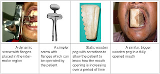
Mouth Opening Device in SMF


Immunological ailments.

Outrageous climatic conditions.

Delayed insufficiency to iron and vitamins in the eating regimen.

DIFFERENTIAL DIAGNOSIS:


Oral appearances of scleroderma

Oral appearances of Plummer Vinson disorder (Iron lack Anemia).

Examination:

Finish Hemogram

Toludine blue test

Biopsy :- Incisional biopsy

Immunofluorescent test:

a) Direct b) Indirect

Administration AND PREVENTION:

The treatment of patients with OSF relies upon the level of clinical contribution. On the off chance that the malady is identified at a beginning period, suspension of the propensity is adequate. Most patients with OSMF give moderateto-serious arranging. Direct to-serious arranging of OSF is irreversible. 

Medicinal treatment is symptomatic and gone for enhancing mouth developments.

Not to devour areca nut and other incessant aggravation, for example, hot and zesty sustenance including chiles.

Guidance green verdant vegetables.

Organization of Vit. A, B complex and high protein consume less calories.

Organization of Antoxid OD for 6 – two months.

Organization of Lycored OD for 6 two months.

Keeping up legitimate oral cleanliness.

Supplementing the eating regimen with nourishments rich in vitamins A, B complex, and C and iron.

Swearing off hot liquids like tea, espresso.

Swearing off liquor.

Utilizing a dental specialist to round off sharp teeth and concentrate third molars.

SURGICAL MANAGEMENT:

Surgical treatment is shown in patients with extreme conditions. These incorporate:-

Basic extraction of the stringy groups: Excision can bring about contracture of the tissue and intensification of the condition.

Split-thickness skin joining following respective temporalis myotomy or coronoidectomy: Trismus related with OSF might be because of changes in the temporalis ligament auxiliary to OSF; subsequently, skin unions may assuage.

Nasolabial folds and lingual pedicle folds: Surgery performed just in patients with OSF in whom the tongue isn't included.

PHYSIOTHERAPY MANAGEMENT: 


Mouth Opening Exercise In SMF


Muscle extending practices for the mouth might be useful to anticipate advance restrictions of mouth opening strong mouth opening has been attempted with mouth choke and non-cyclic surgical screw.

Diathermy: Microwave diathermy appears to be better than short wave, in light of the fact that specific warming of juxtaepitheliel connective tissue is conceivable it acts by physio fibrinolysis of groups.

Ultrasound: Ultra sound selectivity bring the temperature up in some all around aggregated territories. Ultrasound turns out to be an effective profound warming methodology.


Monday, 1 January 2018

Wryneck ( Torticollis ) : Physiotherapy Treatment

TORTICOLIS

Congenital Wryneck
Congenital Wryneck


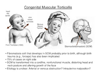
Torticolis is a condition (otherwise called 'wryneck') in which the infant's head is tilted. The head frequently pivots towards one shoulder and tilts away to the contrary side. The term 'intrinsic' is additionally at some point utilized while depicting torticollis. This implies it is available at or not long after birth. Infants treated right on time with physiotherapy programs for the most part react well to treatment.

Torticollis is a side effect identified with turning or twisting of the neck. A wide range of causes are conceivable. In babies, torticollis for the most part comes about because of damage amid work and conveyance or the newborn child's position in the womb. Less regularly, it is caused by birth surrenders. In more seasoned youngsters, torticollis may come about because of wounds to the neck muscles, regular contaminations, or different causes.

Agonizing fits of the neck muscles may happen.


Sternocleidomustoid Muscle


Different indications might be available, contingent upon the reason. For instance, there might be a delicate lymph hub (organ) if the reason is disease.

Sorts:

Inherent torticolis.

Obtained torticolis.


Life systems:

The ordinary physiologic scope of pivot of the map book on the hub is 25-53 degrees to either side. The transverse tendon is the essential stabilizer of the atlantoaxial joint and averts unnecessary foremost movement of the chart book on the pivot. It reaches out behind the lairs, between the average segments of the parallel masses of C1. The combined alar tendons go about as optional stabilizers to anticipate front move. The alar tendons reach out from the parallel part of the caves tip to the average part of the occipital condyles, with a lower partition joining to the average part of the sidelong masses of C1.

The sternocleidomastoid muscle has a sternal and clavicular head. The sternal head is coordinated from the manubrium sterni superiorly, along the side and posteriorly and the clavicular from the average third of the clavicle vertically upward. It races to the mastoid procedure. It empowers an ipsilateral horizontal flexion and a contralateral revolution. The muscle expands the upper piece of the cervical spine and flexes the lower part.

Acquired Torticollis


ETIOLOGY :

Solid in over 80% of the cases. Sorts strong torticollis

  • - Fibromatosis colli: torticollis with obvious mass in the SCM;
  • - Tightness of the SCM without a clear mass;
  • Postural torticollis with neither mass or snugness.
  • Birth injury: aspect separation, tears in the sternocleidomastoid muscle
  • Intrinsic inconsistencies of the craniovertebral intersection: occipitoatlantal combination or Klippel-Feil disorder.
  • Sternocleidomastoid tumor.
  • Visual anomalies.
  • Intrauterine mechanical variables

CAUSES OF WRYNECK :

Sitting or dozing in an irregular position without satisfactory neck bolster.

Poor stance when taking a gander at a PC screen.

Conveying overwhelming lopsided burdens (for instance, a folder case or shopping pack).

Enabling certain muscles of the neck to be presented to chilly (dozing in a draft).

Manifestations:

The turning of your neck (torticollis) happens when your muscles supporting the neck on one side are agonizing.

The agony is as a rule on one side of your neck and solidness of the muscles around there turns the neck to the other side. You may think that its exceptionally troublesome when you attempt to rectify your neck, because of agony. Sporadically, the agony is amidst your neck.

The torment may spread to the back of your make a beeline for your shoulder. The muscles of your influenced side might be delicate. Weight on specific zones may trigger a 'fit' of these muscles. Development of your neck is limited, especially on one side.


Diagnosis :

An exhaustive neurologic examination ought to be performed, and anteroposterior and sidelong radiographs of the cervical spine ought to be gotten. A CT sweep or MRI of the head and neck is important for any patient with relentless neck torment or with neurologic signs and indications.


Medicines:

Agony executioner are regularly useful. for example,

Paracetamol at quality is frequently adequate.

Calming painkillers.

A more grounded torment executioner such codeine.

A muscle relaxant, for example, diazepam.

Different medicines, for example,

Rest.

A decent stance.

A firm supporting pad.

Warmth pack.


PHYSIOTHERAPY TREATMENTS AND EXERCISES : 

Stretching Exercise for Wryneck


Situating.

Delicate scope of movement practices for neck.

Extending of sternocleido mastoid muscle.

Strengthening works out.

Exercises to support dynamic head development.

Visual following.

Horizontal head tilt.

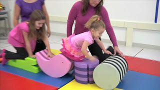
Treatment ball works out.

Side sitting activities.

Hands and knees.

Stooping to standing.

Helped rolling.

Proped sidelying.

Torticollis treatment at home for babies:


Exercise in Torticollis
Exercise In Torticollis


The best technique for torticollis treatment is to urge your infant to hand his or her head over the two bearings. This will relax tense neck muscles and fix the free ones. Here are a few activities to attempt:

At the point when your child needs to eat, offer the jug or your bosom in a way that urges your infant to get some distance from the favored side. (Utilize your tyke's want to eat to support him or her along!)

When putting your infant down to rest, position him or her to confront the divider. Since babies like to watch out onto the room, your infant will effectively get some distance from the divider and this will extend the fixed muscles of the neck.

Amid play, draw your child's consideration with toys and sounds to make him or her hand over the two bearings.


Other Related Article :

Erb's Palsy
Erb's Palsy
 

Monday, 11 December 2017

Erb's Palsy And Physiotherapy :

Erb's palsy And Physiotherapy Treatment : 


Erb's Palsy From Front And Back Side


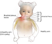
Erb's palsy also called Erb's Duchenne palsy is a paralysis of the arm(Upper Limb). This injury is caused mainly Due to injury to the upper group of the arm's main nerves, Mainly the injury of the upper trunk C5–C6 nerves root. These Nerve Root form part of the brachial plexus, Forming the ventral rami of spinal nerves C5–C8 and One thoracic nerve T1. These injuries Occurs most commonly, but not exclusively, from shoulder dystocia during a difficult birth. Depending on the nature of the damage, the paralysis can either resolve on its own over a period of months, necessary  Physiotherapy Treatment or Severe Injury May require surgical Intervention.

Nerve Root Explanation From Cervical Area


The paralysis can be partial or complete; the damage to each nerve can range from bruising to Complete Tear. The most commonly involved Nerve root is C5 (aka Erb's point: the union of C5 - C6 roots) as this is mechanically the furthest point from the force of traction, therefore, the first/most affected Nerve Root. Erb–Duchenne palsy presents as a lower motor neuron Injury with sensibility Loss and vegetative phenomena.

Erb's Palsy
Infant Nerve Root And Brachial Plexus


The most commonly involved nerves are the suprascapular nerve, musculocutaneous nerve, and the axillary nerve.

The signs of Erb's Palsy include loss of sensation in the arm and paralysis or wekness of the deltoid, biceps, and brachialis muscles. "The position of the limb, under such conditions, is by : the arm hangs by the adducted and is rotate internally ; the forearm is in pronation and exntension position. Sholder Abduction, elbow Flexion And Supination is lost Mainly. The resulting Condition Look's Like Postion Also Called "waiter's tip Hand ".


Brachial Plexus
Brachial Plexus


If this injury occurs at early age May Leads to affect development (e.g. as a neonate or infant), it often leaves the patient with Delayed growth in the affected arm with everything from the shoulder through to the fingertips smaller than Compare to Normal arm. This also leaves the patient with Delayed muscular, Nervous & circulatory development. The Delayed of musculer development May leads to the arm being much weaker than a Normal one, and less articulation, with many patients unable to lift the arm above shoulder height, as well as leaving many with a Muscle contracture.


Which are the Cause of Erb's Palsy ?

Cause Of Erb's Palsy


  • Congenital
  • Dystocia ( Difficult ChildBirth-Labor)
  • Fracture At Clavicle to Neonates.
  • Any age following trauma to the head and shoulder.

Waiter's Tip Hand Position


How Diagnosis is done in Erb's Palsy ?

Examine The Patient's Arm Position Like Adducted From Sholder, Extended From Eblob Joint And Pronated Position With Weakness or Paralysis Of Deltoid, Brachialis,Biceps Most Commonly.
Further Investigation Is By EMG/NCV Reports Or By MRI Accordingly.


Prognosis Of Erb's Palsy


Treatment :

Treatment in Erb's Palsy


Some babies recover on Gradually With Physiotherapy Treatment however, Patient some may require specialist intervention or Surgical Procedure According To Injury.

Neonatal/pediatric neurosurgery is often required for avulsion Injury. Lesions may heal Naturally Over Time and function Gradually return With Help Of Exercise Therapy.

Physiotherapeutic care is required Mainly to restore muscle Function. Although range of motion is recovered in many children under one year in age, individuals who have not yet healed after this point will rarely gain full function in their arm and may develop Deformity.


The three most common treatments for Unrecoverable Erb's Palsy are :

1. Nerve transfers (usually from the opposite arm or limb)
2. Sub Scapularis releases and Latissimus Dorsi Tendon Transfers.


Physiotherapy Treatment :

Physiotherapy Treatment In Erb's Palsy


Assessment Of Patient Mainly Muscle Chart Of Whole Upper Limb And Range Of Motion And RD Test are required.
Accordingly assessment Physiotherapy Treatment Plan are carried out And Monitoring Progress Report With SD Curve At Every 10 Days Helps Recovery Process Going On.

According to Muscle Chart Strengthening Exercise, Electrical Stimulation, Passive Movement Or Active Assisted Exercise Are Design.
Home Exercise Are Teached To Patient's Relative And Deformity Correction Position And Splinting Training Are Also Required.
Mainly Aeroplane Splint Commonly Used But It May Be Vary According To Condition.


Splinting in Erb's Palsy

 
Aeroplane Splint
Aeroplane Splint

Saturday, 17 August 2013

Scoliosis

SCOLIOSIS

IT'S A SIDEWAYS CURVATURE OF THE SPINE. THERE ARE TWO TYPES OF CURVATURE STRUCTURAL AND NON-STRUCTURAL SCOLIOSIS.

STRUCTURAL SCOLIOSIS IS A PERMANENT DEFORMITY. AND NON-STRUCTURAL SCOLIOSIS IS MOBILE AND TRANSIENT.

IT'S A LATERAL CURVATURE OF THE SPINE. THIS IS CALLED THE PRIMARY CURVE. THE SPINE ABOVE AND BELOW THE PRIMARY CURVE UNDERGOES COMPENSATORY CURVES TO THE OPPOSITE SIDE. LATERAL ROTATION OF THE VERTEBRAE ALSO OCCURS.

TREATMENT

TREATMENT CONSISTS OF OPERATIVE AND NON-OPERATIVE TREATMENT.
NON-OPERATIVE TREATMENT CONSISTS OF EXERCISE AND BRACES.