Showing posts with label Paralysis. Show all posts

Sunday, 5 May 2024

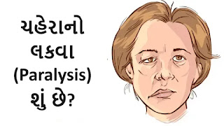
ચહેરાનો લકવા (Paralysis) શું છે?

ચહેરાનો લકવા
ચહેરાનો લકવા

ચહેરાનો લકવો એ એક એવી સ્થિતિ છે જેમાં ચહેરાના સ્નાયુઓને નુકસાન થાય છે, જેના કારણે ચહેરાનો એક ભાગ અથવા સંપૂર્ણ ચહેરો નબળો પડી જાય છે અથવા હલવાનું બંધ કરી દે છે. આ સ્થિતિ ઘણી બધી બાબતોને કારણે થઈ શકે છે, જેમ કે:

  • સ્ટ્રોક: સ્ટ્રોક એ મગજમાં લોહીનો પ્રવાહ અવરોધિત થવાથી થાય છે, જેના કારણે મગજના કોષોને નુકસાન થાય છે અથવા મૃત્યુ પામે છે. મગજનો тот, જે ચહેરાના સ્નાયુઓને નિયંત્રિત કરે છે, તે સ્ટ્રોકથી અસરગ્રસ્ત થઈ શકે છે, જેના કારણે ચહેરાનો લકવો થઈ શકે છે.
  • બેલનું પેરેલિસિસ: બેલનું પેરેલિસિસ એ ચહેરાના સ્નાયુઓને નિયંત્રિત કરતી ચેતાને અસર કરતી એક રહસ્યમય સ્થિતિ છે. તે ચહેરાના એક બાજુ પર અચાનક લકવો લાવી શકે છે.
  • ટ્રોમા: ચહેરા પર ઈજા થવાથી ચહેરાના સ્નાયુઓને નુકસાન થઈ શકે છે, જેના કારણે લકવો થઈ શકે છે.
  • ગાંઠો: મગજ અથવા ચહેરાના સ્નાયુઓમાં ગાંઠો ચહેરાના સ્નાયુઓને નુકસાન પહોંચાડી શકે છે, જેના કારણે લકવો થઈ શકે છે.
  • સંક્રમણ: કેટલાક ચેપ, જેમ કે લાઇમ રોગ, ચહેરાના સ્નાયુઓને નિયંત્રિત કરતી ચેતાને અસર કરી શકે છે, જેના કારણે લકવો થઈ શકે છે.

ચહેરાના લકવાના લક્ષણોમાં શામેલ હોઈ શકે છે:

  • ચહેરાના એક બાજુનો સુન્નતા અથવા નબળાઈ
  • આંખ બંધ કરવામાં અસમર્થતા
  • ઢીલું પડેલું મોઢું
  • લાળ ટપકવું
  • ખાવા-પીવામાં તકલીફ
  • બોલવામાં તકલીફ

જો તમને ચહેરાના લકવાના કોઈપણ લક્ષણો અનુભવાય, તો તરત જ તબીબી સહાય લેવી મહત્વપૂર્ણ છે. ચહેરાના લકવાની સારવાર કારણ પર આધાર રાખે છે. સ્ટ્રોકના કિસ્સામાં, લોહીના ગંઠાઓને તોડવા અથવા દૂર કરવા માટે દવાઓ અથવા સર્જરીનો ઉપયોગ કરી શકાય છે. 

બેલના પેરેલિસિસના કિસ્સામાં, સ્ટીરોઇડ દવાઓનો ઉપયોગ સોજો અને બળતરા ઘટાડવા માટે કરી શકાય છે. ટ્રોમા, ગાંઠો અથવા ચેપના કિસ્સામાં, અંતર્ગત કારણની સારવાર કરવાની જરૂર પડશે.

Monday, 11 December 2017

Erb's Palsy And Physiotherapy :

Erb's palsy And Physiotherapy Treatment : 


Erb's Palsy From Front And Back Side


Erb's palsy also called Erb's Duchenne palsy is a paralysis of the arm(Upper Limb). This injury is caused mainly Due to injury to the upper group of the arm's main nerves, Mainly the injury of the upper trunk C5–C6 nerves root. These Nerve Root form part of the brachial plexus, Forming the ventral rami of spinal nerves C5–C8 and One thoracic nerve T1. These injuries Occurs most commonly, but not exclusively, from shoulder dystocia during a difficult birth. Depending on the nature of the damage, the paralysis can either resolve on its own over a period of months, necessary  Physiotherapy Treatment or Severe Injury May require surgical Intervention.

Nerve Root Explanation From Cervical Area


The paralysis can be partial or complete; the damage to each nerve can range from bruising to Complete Tear. The most commonly involved Nerve root is C5 (aka Erb's point: the union of C5 - C6 roots) as this is mechanically the furthest point from the force of traction, therefore, the first/most affected Nerve Root. Erb–Duchenne palsy presents as a lower motor neuron Injury with sensibility Loss and vegetative phenomena.

Erb's Palsy
Infant Nerve Root And Brachial Plexus


The most commonly involved nerves are the suprascapular nerve, musculocutaneous nerve, and the axillary nerve.

The signs of Erb's Palsy include loss of sensation in the arm and paralysis or wekness of the deltoid, biceps, and brachialis muscles. "The position of the limb, under such conditions, is by : the arm hangs by the adducted and is rotate internally ; the forearm is in pronation and exntension position. Sholder Abduction, elbow Flexion And Supination is lost Mainly. The resulting Condition Look's Like Postion Also Called "waiter's tip Hand ".


Brachial Plexus
Brachial Plexus


If this injury occurs at early age May Leads to affect development (e.g. as a neonate or infant), it often leaves the patient with Delayed growth in the affected arm with everything from the shoulder through to the fingertips smaller than Compare to Normal arm. This also leaves the patient with Delayed muscular, Nervous & circulatory development. The Delayed of musculer development May leads to the arm being much weaker than a Normal one, and less articulation, with many patients unable to lift the arm above shoulder height, as well as leaving many with a Muscle contracture.


Which are the Cause of Erb's Palsy ?

Cause Of Erb's Palsy


  • Congenital
  • Dystocia ( Difficult ChildBirth-Labor)
  • Fracture At Clavicle to Neonates.
  • Any age following trauma to the head and shoulder.

Waiter's Tip Hand Position


How Diagnosis is done in Erb's Palsy ?

Examine The Patient's Arm Position Like Adducted From Sholder, Extended From Eblob Joint And Pronated Position With Weakness or Paralysis Of Deltoid, Brachialis,Biceps Most Commonly.
Further Investigation Is By EMG/NCV Reports Or By MRI Accordingly.


Prognosis Of Erb's Palsy


Treatment :

Treatment in Erb's Palsy


Some babies recover on Gradually With Physiotherapy Treatment however, Patient some may require specialist intervention or Surgical Procedure According To Injury.

Neonatal/pediatric neurosurgery is often required for avulsion Injury. Lesions may heal Naturally Over Time and function Gradually return With Help Of Exercise Therapy.

Physiotherapeutic care is required Mainly to restore muscle Function. Although range of motion is recovered in many children under one year in age, individuals who have not yet healed after this point will rarely gain full function in their arm and may develop Deformity.


The three most common treatments for Unrecoverable Erb's Palsy are :

1. Nerve transfers (usually from the opposite arm or limb)
2. Sub Scapularis releases and Latissimus Dorsi Tendon Transfers.


Physiotherapy Treatment :

Physiotherapy Treatment In Erb's Palsy


Assessment Of Patient Mainly Muscle Chart Of Whole Upper Limb And Range Of Motion And RD Test are required.
Accordingly assessment Physiotherapy Treatment Plan are carried out And Monitoring Progress Report With SD Curve At Every 10 Days Helps Recovery Process Going On.

According to Muscle Chart Strengthening Exercise, Electrical Stimulation, Passive Movement Or Active Assisted Exercise Are Design.
Home Exercise Are Teached To Patient's Relative And Deformity Correction Position And Splinting Training Are Also Required.
Mainly Aeroplane Splint Commonly Used But It May Be Vary According To Condition.


Splinting in Erb's Palsy

 
Aeroplane Splint
Aeroplane Splint

Monday, 20 November 2017

Carpel Tunnel Syndrome And Exercise :


Carpel Tunnel Syndrome: Overview And Physiotherapy :

Carpel Tunnel Syndrome Introduction


Carpel Tunnel Syndrome is tingling numbness, weakness Of Palm Muscle, and Parasthesia in your hand in the area of the Median Nerve because of pressure on the median nerve in your wrist.

The median nerve and several tendons run from your forearm to your hand through a small space in your wrist called the carpal tunnel.


Causes Of carpal tunnel syndrome?

About Carpel Tunnel:


Median Nerve Entering Palm Through Carpel Tunnel


The carpal tunnel is a narrow passage in the wrist Joint , about an inch wide. The floor and sides of the tunnel are formed through small wrist bones called carpal bones.
The roof of the tunnel is a strong band of connective tissue called the transverse carpal ligament. Because these boundaries are very rigid and tight , the carpal tunnel has little capacity to "stretch enough " or increase in size.
The median nerve goes down the arm and forearm, passes through the carpal tunnel at the wrist, and goes into the hand. The nerve provides feeling/control Movement in the thumb and index, middle, and ring fingers. The nerve also controls the muscles around the base of the thumb.
The nine tendons that bend the fingers and thumb also travel through the carpal tunnel. These tendons are called flexor tendons.


Carpal tunnel syndrome occurs when the tunnel becomes narrowed or when tissues surrounding the flexor tendons become swell, Giving Extra pressure on the median nerve.

Pressure on the median nerve causes carpal tunnel syndrome. This pressure can come from swelling or anything that makes the carpal tunnel smaller. Many things can cause this swelling, including:


  • Illnesses such as hypothyroidism, rheumatoid arthritis, and diabetes.
  • overactivity (Making the same hand movements over and over, especially if the wrist is bent down )
  • Pregnancy/Obesity.
  •  Injury Around Wrist Like Fracture, Other Injury.
Symptoms?

Symptoms In Carpel Tunnel Syndrome


Carpal tunnel syndrome can cause tingling numbness, weakness of Palm Muscle Supplied By Median Nerve or pain in the fingers or handAnd Parasthesia. Some people may have Refer pain in their arm between their hand and their elbow.

Symptoms most often occur in the thumb, index finger, middle finger, and half of the ring finger. If you have problems with your other fingers but your little finger is fine, this may be a sign that you have carpal tunnel syndrome. A Ulner nerve gives feeling to the little finger.

You may first notice symptoms at night. You may be able to get relief by shaking your hand, Or In Wrist Mid Extension Position.


Diagnosis?

Your doctor will ask if you have any health problems-such as arthritis, hypothyroidism, or diabetes-or if you are pregnant. He or she will ask if you recently injury Near your wrist, arm, or neck. Your doctor will Take History About your daily routine and any recent activities that could have injury your wrist.

During the exam, your doctor will check the Sensation, Muscle Power , and Positioning of your neck, shoulders, arms, wrists, and hands. Your doctor may suggest tests, such as blood tests or nerve tests Like EMG Or NCV Specially For Median Nerve.


Treatment?


Treatment In Carpel Tunnel Syndrome

Mild symptoms usually can be treated with home care. You can:
  • Avoid activities that cause numbness, pain.
  • Rest your wrist enough between activities.
  • Ice your wrist for 10 to 15 minutes 1 or 2 times an hour.
  • Try taking anti-inflammatory drugs (NSAIDs) to relieve pain and reduce swelling.
  • Wear a wrist splint at night. This takes the pressure off your median nerve.
  • Consult Physiotherapist Or Orthopaedic As Early As Possible.

Physiotherapy Exercise :


Common Wrist Flexor Muscle Stretching Position


Generally, Muscle Supplied By Median Nerve Are Require Strenthening Exercise, So According Assessment Of Muscle Of Palm, And Then Active Movement, Or With Electrical Stimulation, And For Relieving Pain Use Ultrasound, Or Infrared.
Stretching Exercise Of Common Wrist Flexors Is Too Important Without Affecting Pain. 


Splinting :


Neutral Position in Pain Relieving Position is Ideal For Splinting And Night Use Of Splint Lead To Relieve Pressure On Median Nerve.


Splint In Carpel Tunnel Syndrome

 As Soon As you starting treatment, The Symtoms Gradually Down And Relief From Pain And Other Symptoms and preventing long-term damage to the nerve.

Medicine For Other Cause Like Diabetes Or Hyperthyroidism Properly.

Surgery is an optional. But it's usually used only when symptoms are Not Improving that you can't work or do other things even after Few weeks to months of Physiotherapy treatment.


Prevention is Better Than Cure :


To keep carpal tunnel syndrome from coming Again, take care of your basic health. Stay at healthy And Fit. Don't smoke. Exercise to stay strong and flexible. If you have a long-term health problem, such as arthritis or diabetes, follow your doctor's advice for keeping your condition under control.

You can also try to take good care of your wrists and hands:

  • Do Wrist Muscle Stretching Exercise At Regular Interval.
  • Try to keep your wrist in a neutral position.
  • Use your whole hand-not just your fingers-to hold objects.And Also Alternate Hands.
  • When you type, keep your wrists straight, with your hands a little higher than your wrists. Relax your shoulders when your arms are at your sides.
  • If you can Alternate hands Regularly when you repeat movements.
  • Take Enough Rest In Between Activity.
  
Related Article :

Piriformis Stretching

Thursday, 19 September 2013

GBS ( Gullian Barre Syndrome )

What is Guillain-Barre Syndrome (GBS)?

Guillain-Barre Syndrome (GBS), pronounced "ghee-YAN bah-RAY," is a rare neurological disorder in which the body's immune system mistakenly attacks the peripheral nerves. This condition can lead to weakness, numbness, and tingling sensations, and in severe cases, it can cause paralysis. GBS typically develops rapidly and can progress over the course of a few weeks.


  PREDISPOSING FACTORS:

AGE: COMMON BETWEEN 15 TO 25 YEARS.
SEX: COMMON IN FEMALES
INFECTION: VIRAL IN THE FORM OF EPSTEIN BARR VIRUS.
VACCINATION: RABIES, TYPHOID, TETANUS, OR INFLUENZA.
IDIOPATHIC:

Sign and Symptoms

Some common signs and symptoms of Guillain-Barre Syndrome include:

Muscle weakness, often starting in the legs and spreading to the arms.
Tingling or numbness in the extremities.
Unsteady gait or difficulty walking.
Loss of muscle reflexes.
Pain, often described as aching or cramping.
Difficulty with facial movements, speaking, chewing, or swallowing.
Breathing difficulties in severe cases.

The exact cause of GBS is not fully understood, but it is often preceded by an infection, most commonly respiratory or gastrointestinal infections. It is believed that the body's immune response to the infection triggers the autoimmune reaction that leads to the damage of peripheral nerves.

Diagnosis of GBS typically involves a medical history, physical examination, and various tests such as nerve conduction studies and lumbar puncture to examine cerebrospinal fluid. Early diagnosis and prompt treatment are essential to manage the condition effectively and minimize potential complications.

Treatment

Treatment for GBS often involves hospitalization to monitor the progression of the condition and to provide supportive care. Intravenous immunoglobulin (IVIG) and plasmapheresis are common treatments used to reduce the severity of GBS by modulating the immune response. With appropriate care, most people with Guillain-Barre Syndrome can recover over time, although the recovery period may be lengthy, and some individuals may experience residual weakness.

It's important to consult a healthcare professional if you suspect Guillain-Barre Syndrome or are experiencing symptoms. This condition can be serious, and early intervention is crucial for the best possible outcome.

Tuesday, 17 September 2013

Bell's Palsy And Physiotherapy Treatment

Bell's Palsy And Physiotherapy Treatment

Bell's palsy is a condition that causes paralysis or weakness of one side of the facial muscles. It may cause due to inflammation or injury to the facial nerve (7th cranial nerve) which controls facial muscles.

It is also called facial palsy where the loss of facial expression mainly due to facial muscle loss it's a motor function such as unable to close your eye, unable to smile, unable to show teeth, unable to raise your eyebrow are most common symptoms.

However, It can happen at any age, most commonly occurring mainly between 20 to 60. The Bell's palsy was named on scientist Sir Charles Bell (1774-1842), who has long been considered to be the first to describe idiopathic facial paralysis in the early 19th century.

Bell's palsy is mostly recovered with the help of Medical treatment and Physiotherapy exercises within 3 to 4 weeks, however, it may depend upon the severity and symptoms of your condition.

What causes Bell’s palsy?

There are too many reasons for Bell's Palsy where Facial nerves become inflamed or injured in which facial muscles become paralyzed.

The exact cause of nerve palsy is sometimes unknown, However, the few most common causes are as per below :

The viruses and bacteria infections that have been associated with the development of Bell’s palsy are:
  • Exposure of the ear to extreme cold or severe rain
  • Herpes simplex
  • HIV, which damages the immune system
  • Upper respiratory tract infection
  • sarcoidosis, which causes organ inflammation
  • herpes zoster virus, which causes chickenpox and shingles
  • Infection of the ear
  • Epstein-Barr virus, which causes mononucleosis
  • Lyme disease, which is a bacterial infection caused by infected ticks
  • An autoimmune condition is also a causes palsy
  • Unknown cause called Idiopathic cause
If the facial nerve becomes inflamed by infection leads to swelling, which causes pressure on the nerve in the body canal called the Fallopian canal in which the facial nerve passes.
The inflammation of the nerve may reduce blood flow and oxygen to nerve cells. This may lead to Paralysis of one side of facial muscles.
Facial Nerve
Facial Nerve


What are the symptoms of Bell’s palsy?

Symptoms are variable depending upon the severity, causes. There may be partial muscle weakness to complete paralysis of one side of the face. It depends upon nerve damage or compression, if severe nerve damage or compression occurs, there may be complete paralysis, and it takes a longer time to recover.

The symptoms are usually seen suddenly, and you may find them when you wake up in the morning or when you try facial movements such as eating or drinking.

There may be drooping of the corner of the mouth, with difficulty in closing your eyes and raising your eyebrows. It may affect both sides of your face in rare cases.

The most other common symptoms of Bell’s palsy are:
  • Facial weakness or Paralysis
  • drooping of the corner of the mouth
  • Difficulty in the facial expressions, such as smiling, showing teeth, or frowning
  • Creases and skin fold of the face becomes smoothened
  • Rarely speech affected mainly difficulty pronouncing certain words
  • You will see heaviness or numbness of the affected side of the face
  • Facial muscle spasm
  • Pain, swelling near to ear area.
  • Dry eye and mouth
  • altered taste
  • drooling
  • sensitivity to sound
  • difficulty eating and drinking
  • muscle twitches in the opposite side of the face
  • irritation of the eye on the involved side
  • headache
The symptoms can be similar to other serious conditions, such as a stroke or other brain diseases. If you find these symptoms contact your doctor as early as possible for permanent resolution.

What are the risk factors for Bell’s palsy?

Your risk of the vulnerability of Bell’s palsy if you:

  • If you are pregnant
  • If you have uncontrolled diabetes
  • If you have a lung infection
  • If you have a family history of the stroke, Facial palsy related condition
  • If you have High B.P (Hypertension)
  • If you have an ear infection related to any condition
  • If you have any surgery related to your ear
How is Bell’s palsy diagnosed?

Your Doctor is first to examine you to check How facial muscles are working, They also check your medical history, associated with other diseases such as Diabetes, They also ask you have done any surgery or are open to any cold weather.

Following most common tests are used to verify Bell's Palsy, These are:

  • To check Infection, Blood tests are useful.
  • To check diabetes or other associated conditions, a Blood test is also useful.
  • MRI or CT Scan is also useful to check the possibility of stroke or Brain tumor.
  • EMG Test (electromyography) or NCV Test (Nerve conduction test) in which very thin wire electrodes are inserted into the nerve to check the functionality of the facial nerve.
  • To check Lyme disease, a lumbar puncture is also required rarely.
Bell's Palsy Taping
How is Bell’s palsy treated?

Mostly symptomatic Medical treatment and Physiotherapy treatment and exercise help you to recover within a few weeks depending upon the severity of symptoms.

Medical treatment:

To Relieve Pain and Inflammation mostly NSAIDs (Non-Steroidal Anti-Inflammatory Drugs) are Prescribed by the Your Doctors such as Aceclofenac or Diclofenac.
If any Infection is seen in lab tests, accordingly Antiviral or antibacterial are prescribed.
To lubricate your Eye, your doctor gives you eye drops or eye patches.

Home treatment:

A hot pack or a warm, moist towel over your face to relieve pain
Facial massage
To strengthen your weak muscles, physical therapy and exercises for your facial muscles

Physiotherapy Treatment:

Following are the most common principles are used to relieve facial palsy.
  • To Relieve Pain and Inflammation - Infra-red Therapy or Short wave Diathermy
  • Electrical stimulation to strengthen the weak muscle
  • To maintain muscle property - Galvanic current
  • Facial massage
  • Taping or splinting to avoid facial deviation
  • Active assisted or Passive exercise of Facial muscles depends upon grading of muscles
  • Visual feedback exercise
  • Facial care
Are there any complications in Bell’s palsy?

Bell's palsy will completely recover within a few weeks without any complications. Although, few complications may be seen in severe cases of Bell’s palsy.
  • Dryness in the eye on the affected side may lead to infections, ulcers, or difficulty in vision.
  • Synkinesis- in which synergic movement of facial muscles when you try to smile, your eye also closed.
  • Weak facial muscles mean the face has slightly deviated.
Sciatica
Sciatica


Saturday, 7 September 2013

Paraplegia

PARAPLEGIA
PARALYSIS OF BOTH THE LOWER LIMB WITHOUT ANY INVOLVEMENT OF THE UPPER LIMB IS CALLED AS PARAPLEGIA.
CAUSES :
UPPER MOTOR NEURON LESION
  • TUMORS OF FALX CEREBRI
  • THROMBOSIS OF SUPERIOR SAGITTAL SINUS
  • THROMBOSIS OF UNPAIRED ANTERIOR CEREBRAL ARTERY AFFECTING BOTH LEG AREA
  • SUBACUTE COMBINED DEGENERATION OF CORD
  • MULTIPLE SCLEROSIS
  • ACUTE TRANSVERSE MYELITIS
  • MOTOR NEURON DISEASE
  • EPIDURAL ABSCESS
  • RADIATION MYELOPATHY
  • TRAUMA
  • POTT'S PARAPLEGIA
LOWER MOTOR NEURON LESION
  • ANTERIOR HORN CELL LESION IN CONDITIONS LIKE POLIOMYELITIS, SPINAL MUSCULAR ATROPHY AND MOTOR NEURON DISEASE
  • ROOTS LESION LIKE IN CAUDA EQUINA SYNDROME
  • MYASTHENIA GRAVIS
  • PERIPHERAL NERVE LESION
  • SPINA BIFIDA
 CLINICAL FEATURE:
ABSENCE OF REFLEXES
FLACCIDITY
TIGHTNESS CONTRACTURE
OSTEOPOROSIS
URINARY COMPLICATION
DEPRESSION

TREATMENT:
  1. MAINTAINING PASSIVE AND ACTIVE RANGE OF MOTION
  2. CARE OF SKIN 
  3. CARE OF BLADDER
  4. ACTIVE EXERCISES
  5. MANAGEMENT OF SPASTICITY
  6. TRANSFER ACTIVITIES
  7. GAIT TRAINING