Monday, 22 January 2018

OSTEOPOROSIS : Exercise And Diet

OSTEOPOROSIS : 


I T IS THE GENERIC TERM REFERRING TO STATE OF DECREASED MASS PER UNIT OF A NORMALLY MINERALISED BONE DUE TO LOSS OF BONE PROTEINS.
IT IS THE MOST COMMON SKELETAL DISORDER NEXT ONLY TO ARTHRITIS.
CAUSES:
1) DISUSE:  PROLONGED BED REST OR INACTIVITY
                      PROLONGED CASTING OR SPLINTING
                      PARALYSIS,SPACE TRAVEL ETC.
2) DIET:      CALCIUM,PROTEIN LOW IN DIET
                     CHRONIC ALCOHOLISM
3) DRUG WHOSE PROLONGED USE CAUSES OSTEOPOROSIS 
4) IDIOPATHIC
5) GENETIC
6) CHRONIC ILLNESS
7) NEOPLASM
TREATMENT
  • REST,ANALGESIC AND ANTI INFLAMMATORY DRUGS.
  • MUSCLE RELAXANTS AND SUPPORTS LIKE BELTS,COLLAR ETC.
  • HIGH PROTEIN AND CALCIUM DIET
  • BIPHOSPHONATES
  • ACTIVE AND GRADUALLY STRENTHENING EXERCISE


Wednesday, 10 January 2018

Bicipital Tendinitis And Physiotherapy Treatment : Overview

Biceps tendinitis is inflammation of the tendon around the long head of the biceps muscle.
Bicipital Tendinitis


Introduction:

Biceps tendinosis is caused by degeneration of the tendon from athletics requiring overhead motion or from the normal aging process. 

Anatomy Of Bicipital Tendinitis


Inflam-mation of the biceps tendon in the bicipital groove, which is known as primary biceps tendinitis, occurs in 5 percent of patients with biceps tendinitis.
Biceps tendinitis and tendinosis are commonly accompanied by rotator cuff tears or SLAP (superior labrum anterior to posterior) lesions. Patients with biceps tendinitis or tendinosis usually complain of a deep, throbbing ache in the anterior shoulder. Repetitive overhead motion of the arm initiates or exacerbates the symptoms. The most common isolated clinical finding in biceps tendinitis is bicipital groove point tenderness with the arm in 10 degrees of internal rotation. Local anesthetic injections into the biceps tendon sheath may be therapeutic and diagnostic. Ultrasonography is preferred for visualizing the overall tendon, whereas magnetic resonance imaging or computed tomography arthrography is preferred for visualizing.the intraarticular tendon and related pathology. Conservative management of biceps tendinitis consists of rest, ice, oral analgesics, physical therapy, or corticosteroid injections into the biceps tendon sheath. Surgery should be considered if conservative measures fail after three months, or if there is severe damage to the biceps tendon.

Anatomy and Physiology:


The long head of the biceps tendon rises from the supraglenoid tubercle and the superior glenoid labrum.
The proximal portion of the long head of the biceps tendon is extrasynovial but intra-articular.
5 The tendon travels obliquely inside the shoulder joint, across the humeral head anteriorly, and exits the joint within the bicipital groove of the humeral head beneath the transverse humeral ligament.
 The bicipital groove is defined by the greater tuberosity (lateral) and the lesser tuberosity (medial). The biceps tendon is contained in the rotator interval, a triangular area between the subscapularis and supraspinatus tendons at the shoulder (Figure 1). The rotator interval is responsible for keeping the biceps tendon in its correct location.6–8 Because the rotator interval is usually indistinguishable from the rotator cuff and capsule, lesions of the biceps tendon are usually accompanied by lesions of the rotator cuff

SLAP lesions are often present in patients with biceps tendinitis and tendinosis. The anterosuperior labrum and superior labrum are more likely to tear than the inferior portion of the labrum because they are not attached as tightly to the glenoid.9–13 Additionally, certain conditions that affect the glenohumeral joint may also involve the biceps tendon because it is intra-articular. These may include rheumatologic (e.g., rheumatoid arthritis, lupus), infectious, or other types of reactive or inflammatory conditions.

Symtomes:

Patients with biceps tendinitis often complain of a deep, throbbing ache in the anterior shoulder. The pain is usually localized to the bicipital groove and may radiate toward the insertion of the deltoid muscle, or down to the hand in a radial distribution.
This makes it difficult to distinguish from pain that is secondary to impingement or tendinitis of the rotator cuff, or cervical disk disease. Pain from biceps tendinitis usually worsens at night, especially if the patient sleeps on the affected shoulder.
Repetitive overhead arm motion, pulling, or lifting may also initiate or exacerbate the pain.9 The pain is most noticeable in the follow-through of a throwing motion.3 Instability of the tendon may present as a palpable or audible snap when range of motion of the arm is tested.

Rupture of the biceps tendon is one of the most common musculotendinous tears. If the biceps has ruptured, patients will describe an audible, painful popping, followed by relief of symptoms. The anterior shoulder may be bruised, with a bulge visible above the elbow as the muscle retracts distally from the rupture point. Risk factors of biceps rupture include a history of rotator cuff tear, recurrent tendinitis, contralateral biceps tendon rupture, rheumatoid arthritis, age older than 40 years, and poor conditioning.9 If a patient has a feeling of popping, catching, or locking in the shoulder, a SLAP lesion may be present. This usually occurs after trauma, such as a direct blow to the shoulder, a fall on an outstretched arm, or repetitive overhead motion in athletes.

The most common finding of biceps tendon injury is bicipital groove point tenderness.


PHYSICAL EXAMINATION:

Many provocative tests (i.e., Yergason, Neer, Hawkins, and Speed tests) have been developed to isolate pathology of the biceps tendonhowever, because these tests create impingement underneath the coracoacromial arch, it is difficult to rule out concomitant rotator cuff lesions.
The Yergason test requires the patient to place the arm at his or her side with the elbow flexed at 90 degrees, and supinate against resistance18 (Figure 2). The test is considered positive if pain is referred to the bicipital groove.


The Neer test involves internal rotation of the arm while in the forward flexed position16 (Figure 3). If the patient experiences pain, it is a positive sign of impingement syndrome.


During the Hawkins test, the patient flexes the elbow to 90 degrees while the physician elevates the patient's shoulder to 90 degrees and places the forearm in a neutral position19 (Figure 4). With the arm supported, the humerus is rotated internally. The test is positive if bicipital groove pain is present.


Speed test, the patient tries to flex the shoulder against resistance with the elbow extended and the forearm supinated9,20 (Figure 5). A positive test is pain radiating to the bicipital groove. If any of these tests is positive, it indicates that impingement is present, which can lead to biceps tendinitis or tendinosis.


Advantages and Disadvantages of Radiologic Imaging Studies in the Evaluation of Biceps Tendinitis

IMAGING STUDY :
       
Arthrography (used with MRI or CT to visualize the joint capsule and glenoid labrum)
ADVANTAGES :


CT arthrography shows biceps tendon subluxations, ruptures, dislocations, and SLAP lesion
MRI arthrography is preferable for diagnosing biceps lesions and SLAP lesions14 because the agreement between MRI and arthroscopy for biceps lesions is only 37 percent and 60 percent for rotator cuff lesions
DISADVANTAGES
Invasive
Filling of the biceps tendon sheath is unreliable
Sharp images of the tendon may be lost

Ionizing radiation
Bicipital groove view radiography
ADVANTAGES
Shows the width and medial wall angle of the bicipital groove, spurs in the groove, and supertubercular bone spur or ridge
Inexpensive
DISADVANTAGES
Does not show possible intra-articular disorders of the labrum (soft tissue injuries)

MRI
ADVANTAGES
Excellent evaluation of the superior labral complex and biceps tendon
DISADVANTAGES
Partial tears of the biceps tendon are more difficult to detect than complete ruptures
Expensive5
Poorly

Treatment :


CONSERVATIVE:

Biceps tendinitis or tendinitis may respond to analgesia with nonsteroidal anti-inflammatory drugs (NSAIDs).

PHYSIOTERAPY TREATMENT:


Physiotherapy Treatment in Bicipital Tendinitis
 

Ice, rest from overhead activity, or physical therapy.14 Rehabilitation of an athlete's shoulder involves four phases:


Taping Over Biceps Long And Short Head Also Helpful in Early Stage Of Bicipital Tendinitis.

Taping In Bicipital Tendinitis.


rest; stretching exercises of the scapula, rotator cuff, and posterior capsule;
The goal of stretching is to regain a balanced range of motion without stiffness or pain in any position.

strengthening; and a progressively difficult throwing program.
The patient may begin exercises after the shoulder is pain-free.

Biceps Muscle Strenthening Exercise

Friday, 5 January 2018

Trigeminal Neuralgia and Physiotherapy Treatment :

Trigeminal Nerve


              Trigeminal neuralgia is a chronic pain condition that affects the trigeminal nerve, which carries sensation from your face to your brain. If you have trigeminal neuralgia, even mild stimulation of your face — such as from brushing your teeth or putting on makeup — may trigger a jolt of excruciating pain.

You may initially experience short, mild attacks. But trigeminal neuralgia can progress and cause longer, more-frequent bouts of searing pain. Trigeminal neuralgia affects women more often than men, and it's more likely to occur in people who are older than 50.

Because of the variety of treatment options available, having trigeminal neuralgia doesn't necessarily mean you're doomed to a life of pain. Doctors usually can effectively manage trigeminal neuralgia with medications, injections or surgey

Symptom:-


Area Of Trigeminal Nerve
         Illustration showing branches of the trigeminal nerve 
         Trigeminal neuralgia symptoms may include one or more of these patterns:

(1)Episodes of severe, shooting or Tabbing pain that may feel like an electric shock
(2)Spontaneous attacks of pain or attacks triggered by things such as touching the face, chewing, speaking or brushing teeth
Bouts of pain lasting from a few seconds to several minutes
(3)Episodes of several attacks lasting days, weeks, months or longer — some people have periods when they experience no pain
Constant aching, burning feeling that may occur before it evolves into the spasm-like pain of trigeminal neuralgia
(4)Pain in areas supplied by the trigeminal nerve, including the cheek, jaw, teeth, gums, lips, or less often the eye and forehead
Pain affecting one side of the face at a time, though may rarely affect both sides of the face
(5)Pain focused in one spot or spread in a wider pattern


Causes:-

In trigeminal neuralgia, also called tic douloureux, the trigeminal nerve's function is disrupted. Usually, the problem is contact between a normal blood vessel — in this case, an artery or a vein — and the trigeminal nerve at the base of your brain. This contact puts pressure on the nerve and causes it to malfunction.

Trigeminal neuralgia can occur as a result of aging, or it can be related to multiple sclerosis or a similar disorder that damages the myelin sheath protecting certain nerves. Trigeminal neuralgia can also be caused by a tumor compressing the trigeminal nerve.

Some people may experience trigeminal neuralgia due to a brain lesion or other abnormalities. In other cases, surgical injuries, stroke or facial trauma may be responsible for trigeminal neuralgia.

Treatment:-

Treatment In Trigeminal Neuralgia


The first line of treatment is medication.
The drug of choice is carbamazepine (Tegretol™), which eliminates or brings acceptable pain relief in 69 percent of patients.
Baclofen (Lioresal™) is the second drug of choice and may be more effective if used with low-dose carbamazepine.
Other medications that may be effective include pimozide, phenytoin (Dilantin™), capsaicin, clonazepam (Klonopin™) and amitriptyline (Elavil™).

Surgical procedures:-

(1)Percutaneous trigeminal radiofrequency rhizotomy
This procedure selectively destroys pain-causing nerve fibers while preserving touch fibers.
Lesioning techniques include radiofrequency thermocoagulation, glycerol injection and mechanical trauma. They are used for patients who are poor candidates for major surgery.
Complications can include weakness in chewing, facial numbness, changes in tearing or salivation and, less often, corneal ulcers, severe aching pain (anesthesia dolorosa) or meningitis.

(2)Microvascular decompression of the trigeminal nerve
This surgical technique involves microsurgery to move the vessel, causing compression away from the trigeminal nerve.
Relief is often long lived; however the incidence of facial numbness is much less than in selective rhizotomy and anesthesia dolorosa does not occur.
The procedure is best for patients younger than 65 with no significant medical or surgical risk factors.
Possible complications include asceptic meningitis, with head and neck stiffness; major neurological problems, including deafness and facial nerve dysfunction; mild sensory loss; cranial nerve palsy, causing double vision, facial weakness, hearing loss; and, on very rare occasions, postoperative bleeding and death.
Microvascular decompression brings complete relief to 75 percent to 80 percent of patients. The recurrence rate is 5 percent to 17 percent.

Physiotherapy Management:-

*The aims of physiotherapy management:- 
To decrease pain and functional limitation, and to improve quality of life.
Treatments include the use of electro-physical agent to relieve pain during acute onset.
Manual therapy, exercise therapy for Temporomandibular Joint (TMJ) as well
as self-massage for facial muscles can also help to restore patients’ functions.
Transcutaneous electrical nerve stimulation (TENS) currently is one of the
most commonly used forms of electroanalgesia

Interferential Therapy (IFT) :

Interferential therapy (IFT) is another electro-physical modality commonly used
for pain management in clinical situations. IFT is the application of alternating
medium frequency current (4,000 Hz) with amplitude modulated at low
frequency (0–250 Hz). Several theoretical physiological mechanisms such as
the gate-control theory, increased circulation, descending pain suppression,
block of nerve conduction, and placebo have been proposed in the literature to
support the analgesic effects of IFT reducing pain for patients with trigeminal neuralgia by having them received
fifteen sessions of IFT with treatment duration of thirty minutes. The intensity of
the impulse varied according to patient’s tolerance. The results suggested that
IFT could be considered as one of the electro-physical modalities in reducing
pain for trigeminal neuralgia.

Monday, 1 January 2018

Wryneck ( Torticollis ) : Physiotherapy Treatment

TORTICOLIS

Congenital Wryneck
Congenital Wryneck


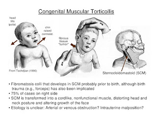
Torticolis is a condition (otherwise called 'wryneck') in which the infant's head is tilted. The head frequently pivots towards one shoulder and tilts away to the contrary side. The term 'intrinsic' is additionally at some point utilized while depicting torticollis. This implies it is available at or not long after birth. Infants treated right on time with physiotherapy programs for the most part react well to treatment.

Torticollis is a side effect identified with turning or twisting of the neck. A wide range of causes are conceivable. In babies, torticollis for the most part comes about because of damage amid work and conveyance or the newborn child's position in the womb. Less regularly, it is caused by birth surrenders. In more seasoned youngsters, torticollis may come about because of wounds to the neck muscles, regular contaminations, or different causes.

Agonizing fits of the neck muscles may happen.


Sternocleidomustoid Muscle


Different indications might be available, contingent upon the reason. For instance, there might be a delicate lymph hub (organ) if the reason is disease.

Sorts:

Inherent torticolis.

Obtained torticolis.


Life systems:

The ordinary physiologic scope of pivot of the map book on the hub is 25-53 degrees to either side. The transverse tendon is the essential stabilizer of the atlantoaxial joint and averts unnecessary foremost movement of the chart book on the pivot. It reaches out behind the lairs, between the average segments of the parallel masses of C1. The combined alar tendons go about as optional stabilizers to anticipate front move. The alar tendons reach out from the parallel part of the caves tip to the average part of the occipital condyles, with a lower partition joining to the average part of the sidelong masses of C1.

The sternocleidomastoid muscle has a sternal and clavicular head. The sternal head is coordinated from the manubrium sterni superiorly, along the side and posteriorly and the clavicular from the average third of the clavicle vertically upward. It races to the mastoid procedure. It empowers an ipsilateral horizontal flexion and a contralateral revolution. The muscle expands the upper piece of the cervical spine and flexes the lower part.

Acquired Torticollis


ETIOLOGY :

Solid in over 80% of the cases. Sorts strong torticollis

  • - Fibromatosis colli: torticollis with obvious mass in the SCM;
  • - Tightness of the SCM without a clear mass;
  • Postural torticollis with neither mass or snugness.
  • Birth injury: aspect separation, tears in the sternocleidomastoid muscle
  • Intrinsic inconsistencies of the craniovertebral intersection: occipitoatlantal combination or Klippel-Feil disorder.
  • Sternocleidomastoid tumor.
  • Visual anomalies.
  • Intrauterine mechanical variables

CAUSES OF WRYNECK :

Sitting or dozing in an irregular position without satisfactory neck bolster.

Poor stance when taking a gander at a PC screen.

Conveying overwhelming lopsided burdens (for instance, a folder case or shopping pack).

Enabling certain muscles of the neck to be presented to chilly (dozing in a draft).

Manifestations:

The turning of your neck (torticollis) happens when your muscles supporting the neck on one side are agonizing.

The agony is as a rule on one side of your neck and solidness of the muscles around there turns the neck to the other side. You may think that its exceptionally troublesome when you attempt to rectify your neck, because of agony. Sporadically, the agony is amidst your neck.

The torment may spread to the back of your make a beeline for your shoulder. The muscles of your influenced side might be delicate. Weight on specific zones may trigger a 'fit' of these muscles. Development of your neck is limited, especially on one side.


Diagnosis :

An exhaustive neurologic examination ought to be performed, and anteroposterior and sidelong radiographs of the cervical spine ought to be gotten. A CT sweep or MRI of the head and neck is important for any patient with relentless neck torment or with neurologic signs and indications.


Medicines:

Agony executioner are regularly useful. for example,

Paracetamol at quality is frequently adequate.

Calming painkillers.

A more grounded torment executioner such codeine.

A muscle relaxant, for example, diazepam.

Different medicines, for example,

Rest.

A decent stance.

A firm supporting pad.

Warmth pack.


PHYSIOTHERAPY TREATMENTS AND EXERCISES : 

Stretching Exercise for Wryneck


Situating.

Delicate scope of movement practices for neck.

Extending of sternocleido mastoid muscle.

Strengthening works out.

Exercises to support dynamic head development.

Visual following.

Horizontal head tilt.

Treatment ball works out.

Side sitting activities.

Hands and knees.

Stooping to standing.

Helped rolling.

Proped sidelying.

Torticollis treatment at home for babies:


Exercise in Torticollis
Exercise In Torticollis


The best technique for torticollis treatment is to urge your infant to hand his or her head over the two bearings. This will relax tense neck muscles and fix the free ones. Here are a few activities to attempt:

At the point when your child needs to eat, offer the jug or your bosom in a way that urges your infant to get some distance from the favored side. (Utilize your tyke's want to eat to support him or her along!)

When putting your infant down to rest, position him or her to confront the divider. Since babies like to watch out onto the room, your infant will effectively get some distance from the divider and this will extend the fixed muscles of the neck.

Amid play, draw your child's consideration with toys and sounds to make him or her hand over the two bearings.


Other Related Article :

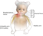
Erb's Palsy
Erb's Palsy