Sunday, 5 May 2024

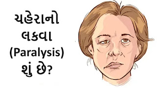
ચહેરાનો લકવા (Paralysis) શું છે?

ચહેરાનો લકવા
ચહેરાનો લકવા

ચહેરાનો લકવો એ એક એવી સ્થિતિ છે જેમાં ચહેરાના સ્નાયુઓને નુકસાન થાય છે, જેના કારણે ચહેરાનો એક ભાગ અથવા સંપૂર્ણ ચહેરો નબળો પડી જાય છે અથવા હલવાનું બંધ કરી દે છે. આ સ્થિતિ ઘણી બધી બાબતોને કારણે થઈ શકે છે, જેમ કે:

  • સ્ટ્રોક: સ્ટ્રોક એ મગજમાં લોહીનો પ્રવાહ અવરોધિત થવાથી થાય છે, જેના કારણે મગજના કોષોને નુકસાન થાય છે અથવા મૃત્યુ પામે છે. મગજનો тот, જે ચહેરાના સ્નાયુઓને નિયંત્રિત કરે છે, તે સ્ટ્રોકથી અસરગ્રસ્ત થઈ શકે છે, જેના કારણે ચહેરાનો લકવો થઈ શકે છે.
  • બેલનું પેરેલિસિસ: બેલનું પેરેલિસિસ એ ચહેરાના સ્નાયુઓને નિયંત્રિત કરતી ચેતાને અસર કરતી એક રહસ્યમય સ્થિતિ છે. તે ચહેરાના એક બાજુ પર અચાનક લકવો લાવી શકે છે.
  • ટ્રોમા: ચહેરા પર ઈજા થવાથી ચહેરાના સ્નાયુઓને નુકસાન થઈ શકે છે, જેના કારણે લકવો થઈ શકે છે.
  • ગાંઠો: મગજ અથવા ચહેરાના સ્નાયુઓમાં ગાંઠો ચહેરાના સ્નાયુઓને નુકસાન પહોંચાડી શકે છે, જેના કારણે લકવો થઈ શકે છે.
  • સંક્રમણ: કેટલાક ચેપ, જેમ કે લાઇમ રોગ, ચહેરાના સ્નાયુઓને નિયંત્રિત કરતી ચેતાને અસર કરી શકે છે, જેના કારણે લકવો થઈ શકે છે.

ચહેરાના લકવાના લક્ષણોમાં શામેલ હોઈ શકે છે:

  • ચહેરાના એક બાજુનો સુન્નતા અથવા નબળાઈ
  • આંખ બંધ કરવામાં અસમર્થતા
  • ઢીલું પડેલું મોઢું
  • લાળ ટપકવું
  • ખાવા-પીવામાં તકલીફ
  • બોલવામાં તકલીફ

જો તમને ચહેરાના લકવાના કોઈપણ લક્ષણો અનુભવાય, તો તરત જ તબીબી સહાય લેવી મહત્વપૂર્ણ છે. ચહેરાના લકવાની સારવાર કારણ પર આધાર રાખે છે. સ્ટ્રોકના કિસ્સામાં, લોહીના ગંઠાઓને તોડવા અથવા દૂર કરવા માટે દવાઓ અથવા સર્જરીનો ઉપયોગ કરી શકાય છે. 

બેલના પેરેલિસિસના કિસ્સામાં, સ્ટીરોઇડ દવાઓનો ઉપયોગ સોજો અને બળતરા ઘટાડવા માટે કરી શકાય છે. ટ્રોમા, ગાંઠો અથવા ચેપના કિસ્સામાં, અંતર્ગત કારણની સારવાર કરવાની જરૂર પડશે.

Wednesday, 6 December 2023

Understanding and Managing Throat Pain During Yawning

Introduction

throat pain on yawning

Yawning is a natural and often involuntary action that occurs throughout the day. While it's commonly associated with tiredness or boredom, many people may experience throat pain during yawning. 

While occasional discomfort is normal, persistent or severe pain might signal an underlying issue. In this article, we'll explore the various causes of throat pain during yawning and discuss effective ways to manage and alleviate the discomfort.

Causes of Throat Pain During Yawning

A. Dry Throat:

Yawning can contribute to throat dryness, leading to irritation and discomfort. Ensuring adequate hydration is crucial in preventing and alleviating dryness. Drinking plenty of water throughout the day can help maintain optimal throat moisture.

B. Muscle Strain:

The muscles in the throat play a significant role during yawning, and strain on these muscles can result in pain. Factors such as overuse, tension, or inflammation may contribute to muscle strain. Taking breaks to rest the voice and practicing relaxation techniques can help reduce muscle tension.

C. Infection or Inflammation:

Throat pain during yawning could be a symptom of infections like strep throat or tonsillitis. Inflammation of the throat tissues may also contribute to discomfort. Seeking medical attention for proper diagnosis and treatment is essential if an infection is suspected.

D. Allergies:

Allergic reactions can irritate the throat, and yawning may exacerbate the discomfort. Identifying and avoiding allergens can help manage throat pain. Antihistamines and allergy management strategies may be recommended by a healthcare professional.

E. GERD (Gastroesophageal Reflux Disease):

Acid reflux can lead to stomach acid flowing back into the throat, causing irritation. Managing GERD through lifestyle changes, such as dietary adjustments and elevating the head during sleep, can alleviate symptoms.

F. Nasal Congestion:

Nasal congestion may result in mouth breathing, leading to throat dryness and discomfort during yawning. Addressing nasal congestion with saline sprays or decongestants can help improve breathing through the nose.

G. Excessive Talking or Singing:

Overuse of the voice, such as excessive talking or singing, can strain the throat muscles. Practicing good vocal hygiene, including warm-up exercises and vocal rest, can prevent muscle fatigue and soreness.

Treatment of Throat Pain During Yawning:

A. Hydration:

Staying adequately hydrated is crucial in maintaining throat moisture. Drinking water throughout the day can prevent dryness and soothe the throat.

B. Throat Lozenges:

Throat lozenges can provide relief by soothing throat irritation. Choosing lozenges with ingredients like menthol or honey can be particularly effective.

C. Avoiding Irritants:

Identifying and avoiding irritants such as smoke or pollutants can contribute to throat health. Creating a throat-friendly environment can help prevent discomfort.

D. Medical Consultation:

If throat pain during yawning persists or is severe, seeking medical advice is important. A healthcare professional can conduct a thorough examination, diagnose the underlying cause, and recommend appropriate treatment.

Conclusion

Throat pain during yawning is a common concern, but understanding its causes and adopting effective management strategies can make a significant difference. Prioritizing throat health, staying hydrated, and seeking timely medical intervention when needed are key steps in ensuring overall well-being. By taking these measures, individuals can minimize discomfort and enjoy the benefits of a healthy and pain-free throat.


Thursday, 25 May 2023

Manual Muscle Testing of Knee Muscles

Manual muscle testing (MMT) is a procedure used to evaluate the strength and function of specific muscles or muscle groups. When it comes to testing the muscles around the knee, MMT can help assess the integrity and strength of the muscles involved in knee movement and stabilization. Here's a general outline of the manual muscle testing process for key muscles around the knee:

Grading scale: The strength is graded on a scale of 0 to 5:

  • 0: No contraction.
  • 1: Flicker contraction, but no movement.
  • 2: Movement with gravity eliminated (i.e., in supine position).
  • 3: Movement against gravity
  • 4: Movement against some resistance
  • 5: Normal strength.

Quadriceps (knee extension):


Manual Muscle Testing of Knee

Position the individual lying on their back with the knee extended.

Place one hand just above the ankle to stabilize the lower leg.

Apply resistance just above the foot, pushing in a downward direction as the individual attempts to straighten their knee.

Gradually increase the resistance while instructing the person to maintain the knee extension.

Assess the strength on a scale from 0 to 5, where 0 represents no muscle contraction and 5 indicates normal strength.

Hamstrings (knee flexion):

Position the individual lying on their stomach with the knee flexed to 90 degrees.

Stabilize the thigh by placing one hand just above the knee joint.

Apply resistance at the heel, pulling in an upward direction as the person attempts to flex their knee against the resistance.

Increase the resistance gradually while instructing the individual to maintain knee flexion.

Use the 0 to 5 scale to evaluate the strength of the hamstrings.

Gastrocnemius and Soleus (ankle plantar flexion):

Position the individual sitting on the edge of a table or chair with their knees flexed to 90 degrees and feet off the ground.

Stabilize the lower leg by holding just above the ankle.

Apply resistance against the ball of the foot, pushing in a downward direction as the person attempts to point their toes downward.

Increase the resistance gradually while assessing the strength.

Use the 0 to 5 scale to rate the strength of ankle plantar flexion.

Remember, manual muscle testing should be performed by a trained professional, such as a physical therapist or healthcare provider, who has knowledge of proper technique and precautions. The procedure may vary depending on the specific muscle being tested and the individual's condition.

Saturday, 14 January 2023

Patellar Tendinitis

What is patellar tendinitis?

Patellar
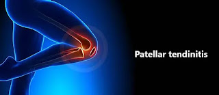
Patellar tendinitis
Patellar tendinitis

tendinitis is an injury to the tendon connecting your kneecap (patella) to your shinbone (tibia). The patellar tendon works with the muscles at the front of your thigh to straighten your knee so that you can kick, run and jump.

Patellar tendinitis is also termed a jumper’s knee. This condition is most common in athletes whose sports involve frequent jumping — sports like basketball and volleyball. However, it is not such that a person who is in sports activity suffer from a condition,, even people who don’t participate in jumping sports can get patellar tendinitis.

Patellar tendonitis condition happens when the patellar tendon becomes inflamed (swollen) or irritated. It is due to overuse or repetitive activity. It typically affects athletes

For most people, treatment of patellar tendinitis starts with physical therapy to stretch and strengthen the muscles around the knee joint.

Patellar tendonitis is also called a:

  • Patellar tendinosis
  • Patellar tendinopathy
  • Jumper’s knee

Patellar tendonitis develops slowly. The condition becomes more severe each time the tendon is overstressed or overused, so it is essential for a person to rest their knee after each injury. Rest will give the body time to heal.

What is a Patellar tendon?

The quadriceps is a muscle whose tendon is located just above the kneecap (patella). The quadriceps tendon connects the quadriceps muscles to the top of the kneecap on the anterior side of the thigh. The patellar tendon is situated just below the kneecap, which connects the kneecap (patella) to the shinbone (tibia). The function of the quadriceps tendon and patellar tendon is to work with the muscles in the front of the thigh to extend or straighten the knee. Therefore, both tendons are important in permitting people to perform activities such as climbing stairs, walking, running, and jumping.

Despite its name, the patellar tendon is truly a ligament. Anatomically, tendons connect a muscle to a bone and ligaments connect two muscles.

The patellar tendon provides stability to the knee joint, holding bones together. It also works in a team with the quadriceps (thigh) muscle and other connective tissues to help in moving. A person couldn’t straighten their knee or jump without it.

What are the Causes of Patellar Tendinitis?

Patellar tendonitis happens when there is more stress on knee tendon tissues, or someone walks too fast, over and over again. Repeated jumping and springing motions stress and strain the bands of patellar tendon tissues. Over time, lots of minor strains and tiny tears make the tendon tissues weaker and sore.

This injury happens slowly over a long time. There may be two main types of activities that damage tendon tissues:

Sudden, increase in activity (how much you’re jumping).

Returning to play at full strength after a long break instead of slowly getting back into your regular routine.

Patellar tendonitis occurs due to repetitive stress on the knee, most often due to overuse in sports or exercise. The repetitive stress on the knee creates tiny tears in the tendon that, over time, inflame and later on weaken the tendon.

Contributing factors can be:

  • Tight leg muscles
  • Uneven leg muscle strength
  • Misaligned feet, ankles, and legs such as Knee Valgus Deformity
  • Obesity
  • Shoes without enough padding
  • Hard playing surfaces
  • Chronic diseases that weaken the tendon

Athletes are more at risk because running, jumping, and squatting put more stress on the patellar tendon. For example, running, or walking can put a force of up to five times more than your body weight on your knees.

If patellar tendonitis remains untreated, a person may have a risk of tendon tear. A large tear of the patella tendon is a serious injury, and a complete tear will separate the patellar tendon from the kneecap. They may hear a tearing or popping sound and feel significant pain when moving.

In a jumper’s knee, it is caused by overuse of your knee joint, such as regular jumping on hard surfaces.

It’s usually a sports-related injury, related to leg muscle contraction and the force of hitting the ground. This force strains the tendon. With repeated stress, a tendon may become inflamed.

Symptoms:

The following are the symptoms:

  • Pain: Pain is the main symptom of patellar tendinitis, usually present between your kneecap and the point where the tendon attaches to your shinbone (tibia).
  • Pain may be a dull type just below the kneecap
  • Pain while jumping, running, or walking
  • Pain when bending or straightening your leg from sitting or standing
  • Kneeling down or getting up from a squatting position can be especially painful.
  • Swelling: The painful tendon may appear thickened or swollen compared to the non-affected tendon on another side.
  • Tenderness around the knee
  • Stiffness: The tendon can often feel unbendable initially in the morning or after physical activity.

Initially, a person may only feel pain in the knee as they begin physical activity or just after an intense workout. Over a long time, the pain progressively worsens and starts to interfere while playing sports like basketball, and volleyball. Later on, the pain interferes with daily and routine movements such as climbing stairs or rising from a chair.

Diagnosis:

It is important not to ignore ongoing pain or discomfort in the knee. Identifying patellar tendonitis early means the condition will be easier and faster to treat, reducing a person’s risk of serious injury.

During the examination, the doctor may apply pressure to parts of your knee to determine where you feel pain. Usually, pain occurs from patellar tendinitis on the front part of your knee, just below your kneecap.

A doctor will ask about:

  • Physical activity
  • what symptoms patient experiencing
  • When the symptoms occur
  • What remedy you’ve tried that eases the pain

The doctor will test the range of motion of the knee by bending and extending the leg.

A doctor may also perform other imaging tests to look at your kneecap and tendon to determine if there is any damage to the tendon or bone. These tests can also help to rule out other possible causes of your pain, such as a fracture or any other condition.

Your doctor may perform:

X-ray: A X-ray to look at the bone to determine whether they have a kneecap fracture or if your kneecap is displaced.

MRI: A MRI to look at the tendon and show any damage to the soft tissue in the knee joint

Ultrasound: An ultrasound to look at the tendon and show any soft tissue damage. Reveal any tears in joint

Complications:

If they do have not medical treatment, patellar tendonitis can worsen. They may damage their tendon more severely, limiting everyday functioning.

Resting legs and stopping activity can be emotionally challenging for athletes, in particular. They may not desire to stop playing, even though it is painful. For professional athletes, patellar tendonitis can be a career-ender if it is left untreated.

Treatment:

Home treatment:

Home remedies

If you have pain in the knee, consider the following:

Pain relievers. Over-the-counter medications such as ibuprofen and naproxen sodium and NSAIDs may provide short-term pain relief.

Avoid activity that causes pain. A person may need to practice their sport less often or temporarily switch to a lower-impact sport. Working or walking with pain can further damage a patellar tendon.

Ice. Apply ice after activity for 10 minutes if that causes pain. Place ice in a plastic bag and wrap the bag in a towel and keep it on the pain area. Or try an ice massage on it. It will reduce swelling and pain.

Medical treatment:

If home remedies do not work, then the person should consult with a doctor for complete treatment. The doctor may provide a pain reliever. It may provide short-term relief from the pain associated with patellar tendinitis.

The doctor may prescribe over-the-counter (OTC) drugs for short-term pain and inflammation reduction.

These can include drugs such as:

  • Ibuprofen
  • Naproxen sodium
  • Acetaminophen

If knee pain is severe, your doctor may give you a corticosteroid injection in the area around the patellar tendon. This treatment is more effective in reducing severe pain. It may also weaken the tendon and possibly make it more likely to rupture. So, it’s important to put considerable thought into this treatment and its associated risks.

Another way of delivering corticosteroid drugs is by spreading the drug over the knee and using a low electrical charge to push it through the skin. This way is called iontophoresis.

Surgical and other procedures

If conservative treatments do not work, the doctor may suggest other therapies, such as:

Corticosteroid injection: An ultrasound-guided corticosteroid injection is injected into the sheath around the patellar tendon may help in relieving pain. But these types of drugs also have complications such as can also weaken tendons and making them more likely to rupture.

Platelet- Rich Plasma injection: This type of injection has been tested in some people with chronic patellar tendon problems. It is hoped the injections might promote new tissue formation and help in healing damage in the tendon.

Oscillating needle procedure. This procedure is performed using local anesthesia. The doctor uses ultrasound imaging to guide a small oscillating needle that cuts away the damaged area while sparing a healthy tendon. This is a fairly new procedure.

Surgery: Surgery is needed in rare cases, if other treatments fail and are successful in relieving pain, the doctor might suggest surgical debridement of the patellar tendon. These procedures can be done through small incisions around the knee.

Traditional surgery involves opening the knee to scrape the damaged knee cap and tendon. More recently, arthroscopic surgery is used for this process. This involves making only four small incisions in the knee, and it has a shorter recovery time, so heals faster.

Recovery time after surgery varies. Some surgical protocols instruct immobility in a cast after your operation. Another strategy for healing suggests that a forceful and immediate rehabilitation exercise program works excellently. In this protocol, people were able to return to high-level activity in 3 months to a year.

Physical Therapy:

The aim of physical therapy is to decrease pain and inflammation. If your pain is unbearable even while you are resting your legs. The doctor may recommend that to support your leg by wearing a brace and using crutches for a while to avoid further damage to the tendon. When movements are relatively pain-free, you can begin physical therapy activities.

Physical therapy can help restore movement gradually as the tendon heals. A physical therapist may also recommend strengthening and stretching exercises people can do at home to do activities without pain in the future.

Additionally, a physical therapist can help in controlling re-injury by finding the cause of the tendonitis. To do this, the doctor can examine a person’s movement patterns and determine why the tendons are being overloaded or overstressed again and again. For example, if a person has weakness in the hips, the knees may take more of the impact when running or jumping, which can cause pain.

In rare cases, untreated tendonitis can lead to a complete tear in the tendon. If this happens, a person may need surgical treatment to reattach the tendon to the bone.

A physical therapy session generally includes:

  • A warm-up period
  • Ice or massage for your knee
  • Stretching exercises
  • Strengthening exercises

Modalities: Modalities such as ultrasound and electrical stimulation are used to ease your knee pain.

Supportive device: A knee brace or taping of your knee may help in reducing pain when you are exercising by keeping the kneecap in place.

A physical therapist will develop an exercise program for you that may include the following exercises:

Stretching

Isometric exercises, where your joint angle and muscle length remain fixed but contractions happen. This exercise helps in relieving pain.

Eccentric exercises are squats performed on a decline board

Flexibility exercises for the thigh and calf muscle

An alternative method of treatments

Exercises and stretches

Doing the exercise in the wrong way, or doing too much too soon, can cause additional damage. People should exercise carefully and do gentle exercises and progress slowly.

Some examples of exercises that may be helpful are given below:

Seated hamstring stretch

Hamstring stretch can be done by sitting on the floor, and stretching one leg straight in front. Keeping the leg straight and toes pointed upwards, gently bend towards the foot. Hold this stretch position for 30 seconds at a time. Can do it 5 times and repeat on both legs.

Quad stretch

Quad stretch
Quad stretch

In a side-lying position, keep the affected knee flexed and extend as possible by using your hand. Hold this position for 30 seconds and 5 times. Slowly remove the force from the leg and repeat on the other side.

Wall Squats

Wall Squats
Wall Squats

Stand with the feet shoulder-width apart while your Back is supported with a wall. Keeping the back straight and neck up, slowly bend from the knees while engaging the glutes. Go as low as you are comfortable. Repeat exercise 8–10 times.

Alternatively or by modifying, a person can try a decline squat by keeping their heels on a small block or thick book. This takes some of the strain off the patellar tendons when performing squatting exercises.

Supine leg lifts

In the supine position, lie on the back with the legs extended up and straight. Slowly lower one leg while keeping the knee in the extended position, then activate the quads to lift the leg. Repeat this exercise on each side.

How to Prevent Patellar Tendinitis?

After an individual has recovered from patellar tendonitis condition, they can take a few steps to prevent future injuries.

The following are prevention for athletes to reduce their risk of injury:

  • Warming up and stretching before starting the exercise
  • Cooling down and stretching after doing exercise
  • Wearing knee support such as a brace when playing sports
  • Knee Exercises to strengthen the leg muscles and support the knees
  • Avoiding jumping, running, and landing on very hard surfaces, such as concrete

Recovery time:

Recovery time for patellar tendonitis varies. It depends on how severely a person has an injury, their overall health and age, and how they treat the condition.

If a person has mild tendonitis, they may be able to return to their normal activities in 3-4 weeks, and in more severe cases, it may take 3 months or more to gets heal.

Some people have pain in their knees even after recovering from patellar tendonitis. Although pain is usually mild and effortless, persistent tendonitis can prevent a person from doing certain activities.

For some athletes, the condition may be chronic. For others, the therapy enables a return to normal functioning and sports.

Important factors in recovery are:

  • Attaching to your rehabilitation program and regularly exercising
  • Resuming your sports activity gradually
  • Paying attention to pain, and resting as required
  • Using Over Counter remedies and ice to help with returning pain

Summary:

Patellar tendonitis is a common knee injury among athletes involving repetitive running and jumping. This stress on the knee damages the patellar tendon, causing pain and inflammation.

The outlook for patellar tendonitis varies based on the severity of the injury. It is such a common occurrence for runners and other athletes.

Patellar tendonitis can develop slowly, so it can be difficult to recognize at first. A person with continued discomfort or pain in the knee should contact a doctor to evaluate their pain.

Resting and supporting by bracing the knee gives a tendon time to heal. In mild cases, patellar tendonitis heals in a few weeks but if the pain continues, a doctor or physical therapist can recommend further treatment options

The best treatment for this condition is to take rest and avoid activities that might aggravate the condition. Doctors may also recommend physical therapy exercises, modifying activities, home treatment-ice and heat, and painkillers.

Tuesday, 30 August 2022

Low Back Pain Cause, Symptoms, Diagnosis, Treatment, Exercise, Ergonomic

Low Back pain
Low Back pain

Low back pain is a general term for pain in your lower back (the part of your spine between your pelvis and your tailbone). It can be acute or chronic. Acute low back pain usually lasts for less than three months, while chronic low back pain lasts for more than three months.

Low back pain is a common condition that affects a large number of people worldwide. For many people, lower back pain can make everyday activities difficult. It's important to understand the causes of this low back pain and find ways to relieve them.

There are several factors that contribute to low back pain, but most cases have an underlying problem. The most common issues include: Lifestyle and the way you take care of yourself, Body Mechanics: Poor muscle strength in your low back, Muscle Imbalance - in which one muscle is stronger than another, Joint abnormalities (such as arthritis), Disc injuries or conditions that can cause pain in the discs between your vertebrae.

Sufferers of low back pain may feel pain in the lower part of their back, though it may radiate down their leg and into the knee. The cause of low back pain is typically a combination of factors such as poor posture that has been incorrectly strengthened through repetitive movements, prolonged sitting at one position, overuse of the spine muscles, abnormal mechanics in the spine or another joint area such as when a person presses their knees together while standing.

The lower back is made up of 5 lumbar vertebrae — L1 through L5 — which are joined together by discs and ligaments. These vertebrae provide stability and support to the spine. The discs act like shock absorbers, cushioning the spine and helping it move naturally when put under stress.

The main function of your lower back is to support your upper body by holding up your spine. When you bend over to pick up something heavy or lean over to reach something on a higher shelf, your legs must use their muscles to help lift you up and your arms must be used to push yourself forward. Your legs are also used for running or other activities that require quick movements. When you sit at a desk all day long or stand for long periods of time, it's important that you have strong muscles in your legs and core so that you can support yourself against gravity.

Low back pain can happen when any part of the spine bends too far forward from its normal position — this can include putting too much weight on one leg while standing or leaning forward from a sitting position

In this post, we cover the causes of low back pain, common symptoms, diagnosis and treatment. We explain what can determine whether you have lumbar spondylytic or herniated disk, and give some suggestions on how to reduce pain in the short term. We also talk about exercise, ergonomics, and diet. The post concludes with a short section on exercises to improve core strength when suffering with low back pain.

  • Section: What is low back pain?
  • Section: Causes of low back pain
  • Section: Symptoms of low back pain
  • Section: Diagnosis of low back pain
  • Section: Treatment of low back pain
  • Section: Exercise for low back pain
  • Section: Ergonomic tips for prevention of low back pain

Takeaway: Taking care of your lower back is important for your health and quality of life.

What Is Low Back Pain?

Low back pain is a common condition that affects a large number of people in worldwide. For many people, lower back pain can make everyday activities difficult. It's important to understand the causes of this low back pain and find ways to relieve them.

Low back pain is usually caused by inflammation or irritation in the muscles, joints, ligaments, tendons, or nerves that support the spine. When these structures become irritated or inflamed, they cause pain and stiffness in the area where they are located. This is also known as referred pain.

What Causes Low Back Pain?

The main cause of low back pain can be due to an injury or illness, or it may be related to other conditions such as arthritis, degenerative disc disease, or chronic fatigue syndrome. In some cases, low back pain may be caused by long-term stress or anxiety. These factors can all affect the way your body responds to injury, illness, or other stressful events.

The most common causes are:

  • Degenerative changes in the bones and joints of the spine
  • Muscle weakness or spasms that limit movement
  • Injuries to muscles and ligaments around the spine, such as broken bones or torn ligaments
  • Injury or strain to lower back muscles
  • Poor posture
  • Lack of exercise and/or physical activity
  • Age-related wear and tear of the spine
  • Stress

other causes are:

Herniated discs: These lumps or bulges in the center of your vertebrae are caused by pressure from the surrounding tissues pushing on them from all sides. Herniated discs are very painful and may also cause sciatica (pain down one or both legs).

Spinal stenosis: This condition occurs when there's too much pressure on one side of your spinal column, which can lead to nerve compression and nerves being pinched off by bone spurs or other structures in the spine

Poor posture: Many people with low back pain have trouble with their posture. This is because they tend to slouch when they're sitting or standing up, which puts extra stress on the muscles and joints in their lower backs. Some people also experience pain when they get up from a sitting position, especially if they have weak abdominal muscles may lead to lumbar lordosis posture (the muscles around your abdomen).

Stress fractures: Fractures that occur as a result of trauma, such as falls or car accidents, are also known as stress fractures.

Stress injuries: These can occur after strenuous activity, such as running up stairs or lifting weights at the gym

Symptoms include:

  • Pain in one or both buttocks and/or thighs
  • Pain radiating down into one or both legs
  • Pain that gets worse with activity
  • Pain that wakes you up at night
  • Tenderness to touch over the spine
  • A feeling of stiffness or tightness in the lower back
  • Difficulty maintaining balance while standing or walking
  • Constipation

Diagnosis:

If you have been experiencing lower back pain for more than 3 weeks, you should see your doctor for an evaluation. An evaluation may include imaging tests such as X-rays or MRIs (magnetic resonance imaging). These images will help doctors determine if there is any damage to your spine and whether or not you have any conditions that would require further treatment.

Your doctor will perform a physical exam.

X-rays show the bones of the spine, discs, and joints that can cause pain if there is damage to these structures. They are usually taken when you stand upright.

MRIs (magnetic resonance imaging) have a stronger pull on the spine than X-rays do, so they can better detect damage to your spine’s soft tissues.

CT (computed tomography) scan

Ultrasonography and Bone density tests also help to diagnose the Low back pain

Low Back Pain Treatment Options

In some cases, treating low back pain with physical therapy can help improve the strength and flexibility of your muscles and ligaments. Other treatments include:

Medication - Pain medications such as acetaminophen (Tylenol) and nonsteroidal anti-inflammatory drugs (NSAIDs) may relieve muscle aches and joint stiffness associated with low back pain. If over-the-counter medications don't provide adequate relief after several weeks, talk with your doctor about using prescription drugs such as ibuprofen

A nerve block can be used to help relieve pain from compression on the nerves in your spine. This may include injections with steroids and anesthetic medications into specific areas along the spine where there is pressure from compressed nerves (epidural space).

Your doctor may recommend that you wear a brace or splint to provide support while your back heals itself over time.

If you have back pain that doesn’t respond to non-surgical treatments, surgery may be recommended.

Low Back pain exercise

The physiotherapy treatment for low back pain includes exercise, physical therapy, and other treatments to relieve discomfort in your back and spine. Exercise may include stretching exercises or strengthening exercises for the muscles around your pelvis and hips that support your spine.

Physical therapy focuses on the movement and function of your spine and muscles. A physical therapist can help with exercises that strengthen the core muscles around your pelvis and back to prevent pain.

Physiotherapy is a back pain treatment that helps reduce pain and restore function. It can help in improving the quality of life by reducing the disability and making sure that you get back to your regular day-to-day activities.

There are a number of exercises that are recommended by physiotherapists to make sure that you recover from back pain. These exercises include strengthening, stretching, aerobic, and core exercises. You can also use heat therapy, hydrotherapy, massage therapy, and acupuncture to reduce the pain.

You will be guided on how to do the exercises and give advice on which methods are more effective. You should not attempt these exercises without consulting a physiotherapist. Massage therapy, acupuncture, heat therapy, hydrotherapy, and exercise programs are also recommended.

Monday, 15 August 2022

Lumbar Lordosis

What is Lumbar Lordosis?

Lumbar Lordosis

Lumbar lordosis is the inward curvature of the lumbar spine. You may notice that some people have a small degree of lordosis (swayback) while others (especially those with very upright sitting postures) have severe lumbar spine curvature. 

It can be caused by a number of conditions such as habitual harmful postures, injury, and fractures, especially after childbirth. The lumbar spine plays a vital role in supporting your body’s weight, and keeping you stable and normal curves serve to distribute mechanical stress incurred as the body is at rest and during movement.

Lumbar lordosis is the inward curve of the lumbar spine. A small degree of lumbar spine curvature is normal while too much lumbar spine curvature is called lordosis (swayback).

The lumbar spine is a part of the trunk that supports the organs in your lower body while controlling muscle movements. In addition, it is used for flexibility and movement energy transmission. A small degree of lordosis is normal while too much lumbar spine curving is called lordosis (swayback).

Lumbar lordosis can be caused by genetic factors, obesity, and pregnancy. The symptoms of lumbar lordosis are related to the severity of the condition. Some people may have no symptoms at all, while others may experience pain in their lower back, numbness in their legs or feet, and difficulty walking.

Symptoms of Lumbar Lordosis

Lumbar lordosis is a condition where the spine curves inwards at the lower back. 

This condition can cause pain in the lower back and hamper mobility.

 Common symptoms of lumbar lordosis include:

  • Lower back pain and/or stiffness
  • It can cause pain and discomfort in the lower back, hips, buttocks, and legs.
  • Hip pain and/or stiffness
  • Tingling or numbness in the feet or toes
  • Pain while sitting for long periods of time
  • Tightness of the hamstrings
  • A feeling of instability or insecurity
  • Difficulty standing up straight
  • Pain during sitting or standing for long periods of time

The symptoms of lumbar lordosis are related to the severity of the condition. Some people may have no symptoms at all, while others may experience pain in their lower back, numbness in their legs or feet, and difficulty walking.

What Causes Lumbar Lordosis?

Lumbar lordosis is the natural inward curve of the spine. It is caused by a combination of genetics, age, and muscle imbalance.

It is a condition in which the back becomes arched or curved. This can happen due to a number of reasons.

 Some causes of lumbar lordosis are:

  • Poor posture
  • sitting for long periods of time
  • working at a desk all-day
  • obesity
  • Some medical conditions such as osteoporosis, scoliosis, and spondylolisthesis

Risk Factor:


The risk factors for lumbar lordosis include 

  • poor posture, 
  • being overweight or obese
  • having weak abdominal muscles
  • Abdominal surgery

Diagnosis Lumbar Lordosis

The condition can be diagnosed by a physical examination, X-rays, and MRI scans. It can also be diagnosed with a special test called the standing flexion test or the standing extension test.

Diagnosis of lumbar lordosis is done through the following steps:

Physical examination: A physical examination can be done to identify the symptoms of lumbar lordosis. The doctor will check for any abnormalities in muscle strength, posture, or gait. The doctor will also examine for any visible signs of injury to the back and spinal cord.

X-ray: An x-ray of the spine can help diagnose lumbar lordosis as well by identifying any structural abnormalities such as scoliosis or spondylolisthesis.

MRI: An MRI scan can also be used to diagnose

How is Lumbar Lordosis Treated?

There are several ways to treat lumbar lordosis.

Medical treatment is mostly symptomatic if you have Pain, the Doctor prescribes you Pain relieving Medicine mainly Non-steroidal anti-inflammatory drugs (NSAIDs) and If your bone becomes weak (osteoporosis) they also give you Calcium with Vitamin d3 combination medicine.

The first option is to use physical therapy to strengthen the muscles around the area. The physiotherapist prescribes you a few exercises and tips that give you a permanent solution and it is a natural way to recover without any side effects.

Exercise

Exercise for Lumbar Lordosis

Exercise is one of the best ways to treat lumbosacral pain. You can try walking, swimming, cycling, running, dancing, yoga, Pilates, weight training, etc. If you have lumbar pain, you should avoid activities that require you to sit or stand for long periods of time.

Stretching exercise

Stretching is a great way to relieve back pain caused by lumber lordosis. Stretching helps to loosen tight muscles and increase flexibility. There are many different types of stretching exercises that can be performed to relieve back pain. 

One example of a stretching exercise is performing a forward bend while holding onto something for balance. Another example would be to lie down on the floor and lift both legs off the ground. These two examples are just a couple of ways to stretch out the back muscles.

Strengthening Exercise:

Strengthening exercises of weak Abdominal muscles improves lumbar curvature and makes your spine more stable and normal.

Massage Therapy

Massage therapy helps relax muscles and reduce tension. It can help improve blood circulation and lymphatic drainage. It can also help increase flexibility and range of motion.

Heat Packs

Heat packs are a great way to relieve muscle tension and promote healing. Heat packs can be placed directly over the affected area to provide relief.

Ice Pack

Ice packs are a great way for patients to relieve back pain. Ice packs are applied directly to the painful area and left on for about 15 minutes. After the ice pack has been removed, the area should be massaged to stimulate blood flow.

Acupuncture

Acupuncture is a traditional Chinese medicine technique that uses fine needles to stimulate points along the meridians of the body. Stimulating these points can have a positive effect on reducing back pain.
Load more 

Chiropractic Care

Chiropractic care focuses on the alignment of the spine. By adjusting the spine, chiropractors can help eliminate nerve interference and restore proper function.

Surgery is the last option you should select if Medical and Physical therapy do not help.

Conclusion: A Guide to Treating Back Pain Caused by Lumber Lordosis

In order to treat lumber lordosis, the patient should perform regular exercises that strengthen their core muscles and help them maintain proper posture. Lordotic posture mostly recovers with the Help of Physiotherapy treatment and exercise, However, you should contact a nearby Physiotherapist or Physician for the Best treatment.

Cervical Spondylosis

Introduction

Neck pain
Neck pain

Spondylosis is a condition that affects your spine or bones and joints in your neck. You can get cervical spondylosis if you have worn out or damaged discs and cartilage in your neck. Cervical spondylosis causes wear and tear on the cartilage and bones of the neck. 

The symptoms of cervical spondylosis include Neck pain, stiffness, chronic headaches, and difficulty walking due to pain from osteoporosis (loss of bone density).

The cervical spine is a part of the spine that connects the head and neck to the rest of the body. It is made up of 7 vertebrae and they are called C1 through C7. The bones in each vertebra can move a little bit, but they are held together by ligaments that hold them together tightly so your head doesn't fall off!

What is cervical spondylosis?

Cervical spondylosis is a degenerative bone disease that affects the vertebrae in the neck. It affects people over the age of 50, with most cases occurring between 50 and 70 years old.

The condition can cause pain and stiffness in your neck, headaches, numbness or tingling sensation in your hands or arms (especially at night), difficulty swallowing (dysphagia), or slurred speech. Some people experience no symptoms at all until they have been diagnosed with cervical spondylosis by their doctor because it often progresses slowly over time before showing signs of illness.

Cervical spondylosis causes

Cervical spondylosis is a condition that causes painful, stiffening of the neck and spine. It can be caused by:

  • Overuse of the neck (injury)
  • Poor posture
  • Genetics
  • Aging
  • Degenerative changes in your joints and bones as you get older

Cervical spondylosis risk factors

Cervical spondylosis is a disease that can affect the joints of your neck, spine, and upper back. Your risk of developing cervical spondylosis increases with age and genetics. If you have a family history of the condition, you are at greater risk of developing it than those who do not have this condition in their family history.

Cervical spondylosis causes pain, stiffness, and limited motion in your neck as well as headaches that may be severe enough to require medication or surgery to relieve them completely from time to time; however, when left untreated this condition has no cure but instead requires constant care by an experienced specialist who understands how the best treatment options will help alleviate symptoms associated with this painful condition!

Cervical spondylosis symptoms

  • Pain in the neck
  • Pain in the shoulder Blade
  • Neck to Shoulder pain
  • Pain in the upper back
  • Pain in the arms
  • Pain in the hands, fingers, and feet.

Cervical spondylosis diagnosis

Cervical spondylosis diagnosis is based on symptoms, other diagnostic tests, and patient history.

Diagnosis of cervical spondylosis is based on patient symptoms, imaging studies (x-rays and MRI), physical examination findings, and a discussion of the results with the doctor. The type of imaging study will vary depending upon what you have been diagnosed with. X-rays are used to look for evidence of damage to bones or joints as well as ligaments and discs in the neck area which could indicate that there may be something wrong with them causing pain in this area such as arthritis or new bone formation around old ones - these things can occur even without any obvious signs being seen by an x-ray so it's important not only to ask yourself if you've had any accidents that caused the injury but also think back over your life history since birth since some people develop problems later down the line which could affect their quality of life later on after they've already started getting worse!

MRI scans use magnetic fields rather than radiation like CTs do; however, both types show up similar images depending upon how strong they're manipulated during processing time."

Treatment:

After your doctor has made a diagnosis, they will discuss treatment options with you. In severe cases, people can undergo surgery to repair the damage. The treatment will depend on the severity of the injury. If a person is experiencing mild symptoms, they may be able to recover with rest, ice, and over-the-counter pain relievers.

Exercises for cervical spondylosis

There are a number of exercises you can do to help ease neck pain. These include:

Active Neck movements such as

  • Neck flexion and extension
  • Neck rotation to each side
  • Chin tuck (pull your chin towards your chest) -
  • Shoulder shrugs (1 second up, 1 second down)
  • Elbow circles forward and backward
  • Lateral flexion of the neck (look left and right, each side)
Isometric Neck exercise

Isometric Neck exercise

Shoulder scapular exercise

Electrotherapy Treatment

Neck traction

TENS (Transcutaneous Electrical Nerve Stimulation)

IFT (Interferential Therapy)

Heat and cold treatments

Massage and soft tissue release or manipulation

Conclusion

Cervical spondylosis is a common condition that affects the joints and discs in your neck. It develops from wear and tears on the cartilage and bones of the neck, which can happen when you're older or if you live a sedentary lifestyle. 

Regular exercise and an Active lifestyle with a Good diet help to prevent Cervical spondylosis.