Monday, 11 December 2017

Erb's Palsy And Physiotherapy :

Erb's palsy And Physiotherapy Treatment : 


Erb's Palsy From Front And Back Side


Erb's palsy also called Erb's Duchenne palsy is a paralysis of the arm(Upper Limb). This injury is caused mainly Due to injury to the upper group of the arm's main nerves, Mainly the injury of the upper trunk C5–C6 nerves root. These Nerve Root form part of the brachial plexus, Forming the ventral rami of spinal nerves C5–C8 and One thoracic nerve T1. These injuries Occurs most commonly, but not exclusively, from shoulder dystocia during a difficult birth. Depending on the nature of the damage, the paralysis can either resolve on its own over a period of months, necessary  Physiotherapy Treatment or Severe Injury May require surgical Intervention.

Nerve Root Explanation From Cervical Area


The paralysis can be partial or complete; the damage to each nerve can range from bruising to Complete Tear. The most commonly involved Nerve root is C5 (aka Erb's point: the union of C5 - C6 roots) as this is mechanically the furthest point from the force of traction, therefore, the first/most affected Nerve Root. Erb–Duchenne palsy presents as a lower motor neuron Injury with sensibility Loss and vegetative phenomena.

Erb's Palsy
Infant Nerve Root And Brachial Plexus


The most commonly involved nerves are the suprascapular nerve, musculocutaneous nerve, and the axillary nerve.

The signs of Erb's Palsy include loss of sensation in the arm and paralysis or wekness of the deltoid, biceps, and brachialis muscles. "The position of the limb, under such conditions, is by : the arm hangs by the adducted and is rotate internally ; the forearm is in pronation and exntension position. Sholder Abduction, elbow Flexion And Supination is lost Mainly. The resulting Condition Look's Like Postion Also Called "waiter's tip Hand ".


Brachial Plexus
Brachial Plexus


If this injury occurs at early age May Leads to affect development (e.g. as a neonate or infant), it often leaves the patient with Delayed growth in the affected arm with everything from the shoulder through to the fingertips smaller than Compare to Normal arm. This also leaves the patient with Delayed muscular, Nervous & circulatory development. The Delayed of musculer development May leads to the arm being much weaker than a Normal one, and less articulation, with many patients unable to lift the arm above shoulder height, as well as leaving many with a Muscle contracture.


Which are the Cause of Erb's Palsy ?

Cause Of Erb's Palsy


  • Congenital
  • Dystocia ( Difficult ChildBirth-Labor)
  • Fracture At Clavicle to Neonates.
  • Any age following trauma to the head and shoulder.

Waiter's Tip Hand Position


How Diagnosis is done in Erb's Palsy ?

Examine The Patient's Arm Position Like Adducted From Sholder, Extended From Eblob Joint And Pronated Position With Weakness or Paralysis Of Deltoid, Brachialis,Biceps Most Commonly.
Further Investigation Is By EMG/NCV Reports Or By MRI Accordingly.


Prognosis Of Erb's Palsy


Treatment :

Treatment in Erb's Palsy


Some babies recover on Gradually With Physiotherapy Treatment however, Patient some may require specialist intervention or Surgical Procedure According To Injury.

Neonatal/pediatric neurosurgery is often required for avulsion Injury. Lesions may heal Naturally Over Time and function Gradually return With Help Of Exercise Therapy.

Physiotherapeutic care is required Mainly to restore muscle Function. Although range of motion is recovered in many children under one year in age, individuals who have not yet healed after this point will rarely gain full function in their arm and may develop Deformity.


The three most common treatments for Unrecoverable Erb's Palsy are :

1. Nerve transfers (usually from the opposite arm or limb)
2. Sub Scapularis releases and Latissimus Dorsi Tendon Transfers.


Physiotherapy Treatment :
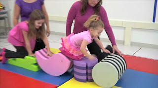
Physiotherapy Treatment In Erb's Palsy


Assessment Of Patient Mainly Muscle Chart Of Whole Upper Limb And Range Of Motion And RD Test are required.
Accordingly assessment Physiotherapy Treatment Plan are carried out And Monitoring Progress Report With SD Curve At Every 10 Days Helps Recovery Process Going On.

According to Muscle Chart Strengthening Exercise, Electrical Stimulation, Passive Movement Or Active Assisted Exercise Are Design.
Home Exercise Are Teached To Patient's Relative And Deformity Correction Position And Splinting Training Are Also Required.
Mainly Aeroplane Splint Commonly Used But It May Be Vary According To Condition.


Splinting in Erb's Palsy

 
Aeroplane Splint
Aeroplane Splint

Friday, 1 December 2017

Parkinson's Disease

Parkinsonism Disease And Physiotherapy Treatment : 


Parkinson's Disease


Presentation:-

Parkinsonism is a clinical disorder portrayed by tremor, bradykinesia, inflexibility, and postural shakiness.

Parkinsonism is any condition that causes a mix of the development anomalies found in Parkinson's ailment —, for example, tremor, moderate development, debilitated discourse or muscle firmness — particularly coming about because of the loss of dopamine-containing nerve cells (neurons). 

Posture In Parkinson's Disease


Life systems:-


Parkinsonism illness (PD) is a degenerative, dynamic issue that influences nerve cells in profound parts of the cerebrum called the basal ganglia and the substantia nigra. Nerve cells in the substantia nigra create the neurotransmitter dopamine and are in charge of handing-off messages that arrangement and control body development. For reasons not yet comprehended, the dopamine-delivering nerve cells of the substantia nigra start to cease to exist in a few people. At the point when 80 percent of dopamine is lost, PD side effects, for example, tremor, gradualness of development, solidness, and adjust issues happen.

Body development is controlled by an unpredictable chain of choices including between associated gatherings of nerve cells called ganglia. Data goes to a focal region of the cerebrum called the striatum, which works with the substantia nigra to send motivations forward and backward from the spinal rope to the mind. The basal ganglia and cerebellum are in charge of guaranteeing that development is done in a smooth, liquid way. 


Stages Of Parkinson's Disease


Phases OF PARKINSONISM :-

STAGE ONE - A Person as a rule has mellow manifestations, for example, tremors or shaking in a limb.Change, for example, poor stance, loss of balance,and strange outward appearances.

STAGE TWO - Symptoms influence the two appendages and the two sides of the body. The individual has for the most part has issue strolling or adjusting, and the failure to finish physical errands turns out to be more clear.

STAGE THREE - Symptoms can be extreme and incorporate the powerlessness to walk straight or stand. there is a detectable abating of physical developments.

STAGE FOUR - The capacity to walk is regularly restricted

STAGE FIVE - tHE PERSON is regularly unfit to deal with herself and will most likely be unable to stand or walk .she may require consistent one on one nursing care.

CAUSES:-

Mind damage

Diffuse Lewy body ailment (a kind of dementia)

Encephalitis

HIV/AIDS

Meningitis

Numerous framework decay

Dynamic supranuclear paralysis

Stroke

Wilson ailment

Different reasons for auxiliary parkinsonism include:

Cerebrum harm caused by anesthesia drugs, (for example, amid surgery)

Carbon monoxide harming

Certain medications used to treat mental scatters or queasiness

Mercury harming and other compound poisonings

Overdoses of opiates

MPTP (a contaminant in some road drugs)

Indications:-

Indications of Parkinson's ailment contrast from individual to individual. They additionally change as the illness advances.

Indications ordinarily start showing up between the ages of 50 and 60. They grow gradually and regularly go unnoticed by family, companions, and even the individual who has them.

The most widely recognized one is tremor.

Tremor. A tremor, or shaking, as a rule starts in an appendage, regularly your hand or fingers. You may see a forward and backward rubbing of your thumb and index finger, known as a pill-moving tremor. One normal for Parkinson's infection is a tremor of your hand when it is casual (very still).

Impeded development (bradykinesia). After some time, Parkinson's sickness may lessen your capacity to move and moderate your development, making basic assignments troublesome and tedious. Your means may end up plainly shorter when you walk, or you may think that its hard to escape a seat. Additionally, you may stall as you endeavor to walk, making it hard to move.

Unbending muscles. Muscle firmness may happen in any piece of your body. The hardened muscles can restrain your scope of movement and cause you torment.

Debilitated stance and adjust. Your stance may wind up plainly stooped, or you may have adjust issues because of Parkinson's malady.

Loss of programmed developments. In Parkinson's malady, you may have a diminished capacity to perform oblivious developments, including flickering, grinning or swinging your arms when you walk.

Discourse changes. You may have discourse issues because of Parkinson's illness. You may talk delicately, rapidly, slur or waver before talking. Your discourse might be all the more a monotone as opposed to with the standard articulations.

Composing changes. It might turn out to be difficult to compose, and you're composing may seem little.

Determination:-

Registered tomography (CT) sweeps of individuals with PD typically seem ordinary.

X-ray has turned out to be more precise in conclusion of the ailment after some time, particularly through iron-delicate T2* and SWI groupings at an attractive field quality of no less than 3T, both of which can exhibit nonattendance of the trademark 'swallow tail' imaging design in the dorsolateral substantia nigra.

TREATMENT:-

Carbidopa-levodopa.

Carbidopa-levodopa mixture.

Dopamine agonists.

MAO-B inhibitors.

Catechol-O-methyltransferase (COMT) inhibitors.

Anticholinergics.

Amantadine.

PHYSIOTHERAPY TREATMENT:- 


Gait Training Exercise

Balancing Exercise

Point OF PHYSIOTHERAPY-

Keep up and enhance levels of capacity and autonomy, which will enhance a man's personal satisfaction

Utilize exercise and development techniques to enhance portability

Rectify and enhance anomalous development examples and stance, where conceivable

Boost muscle quality and joint adaptability

Rectify and enhance stance and adjust, and limit dangers of falls

Keep up a decent breathing example and compelling hack

Instruct the individual with Parkinson's and their carer or relatives

Upgrade the impacts of medication treatment.


Various Exercise In Parkinsonism

Physiotherapy Treatment


TREATMENT PLAN -

Visual prompting – a concentration point to venture over and start step; portions of tape on the floor to start or keep strolling through regions that reason moderating or solidifying

Sound-related prompting – tallying 1-2-3 to start strolling; venturing to the beat of a metronome or particular music at a predefined rhythm to proceed with the cadence of a walk

Consideration – Thinking about making a major stride; influencing a more extensive curve to turn

Proprioceptive prompting – shaking from side to side prepared to start a stage; making one stride in reverse as a sign prepared to then walk advances.


Tuesday, 21 November 2017

Knee Valgus Deformity : Overview:


Knee valgus is as valgus collapse and medial knee displacement. It is characterized by hip adduction and hip internal rotation, usually when in a hips-flexed position (the knee actually abducts and externally rotates)

CAUSES:-


One of the most commonly held theories is that reduced gluteal activity levels allow for a greater degree of hip internal rotation and consequently also tibial rotation and therefore Develop knee valgus.

RISK FACTOR:-

It is most commonly implicated in injuries involving the anterior cruciate ligament (ACL) but it also features as a risk factor in respect of other leg injuries, including patellofemoral pain syndrome, knee osteoarthritis, medial collateral ligament sprains and more general knee cartilage and meniscus damage.

DIAGNOSIS:-


Q Angle Measurement In Knee Valgus
 

The degree of genu valgum can be estimated by the Q angle, which is the angle formed by a line drawn from the anterior superior iliac spine through the center of the patella and a line drawn from the center of the patella to the center of the tibial tubercle.
 

In women, the Q angle should be less than 22 degrees with the knee in extension and less than 9 degrees with the knee in 90 degrees of flexion.
 

In men, the Q angle should be less than 18 degrees with the knee in extension and less than 8 degrees with the knee in 90 degrees of flexion.
 

A typical Q angle is 12 degrees for men and 17 degrees for women.

TREATMENT:-


Splinting In Knee Valgus


It is normal for children to have knock knees between the ages of two and five years of age, and almost all of them resolve as the child grows older. If symptoms are prolonged and pronounced or hereditary, doctors often use orthotic shoes or leg braces at night to gently move a child's leg back into position. If the condition persists and worsens later in life, surgery may be required to relieve pain and complications resulting from severe or hereditary genu valgum. Available surgical procedures include adjustments to the lower femur and total knee replacement (TKR).
Weight loss and substitution of high-impact for low-impact exercise can help slow progression of the condition.

PHYSIOTHERAPY -


Exercise In Knee Valgus


Physiotherapy Treatment


 Activating and developing the arches of the feet,
 Waking up the outer leg muscles (abductors), and
 Learning how to move the inner ankle bone inwards towards the outer ankle bone, and upwards towards the knee.
 

 Strengthening exercise -

                         Side Palnk,
                         Side Step-up,
                         Stationary Lunge Exercise,
                         Lunge Exercise,
                         Rubber Band Squats,
                         Lying Side Leg Lifts,
                         Lying Hip External Rotation,
                         One Legged Wall Push.


Other Knee Joint Deformity Related Article :

          
Knee Varus Deformity Detail

Monday, 20 November 2017

Carpel Tunnel Syndrome And Exercise :


Carpel Tunnel Syndrome: Overview And Physiotherapy :

Carpel Tunnel Syndrome Introduction


Carpel Tunnel Syndrome is tingling numbness, weakness Of Palm Muscle, and Parasthesia in your hand in the area of the Median Nerve because of pressure on the median nerve in your wrist.

The median nerve and several tendons run from your forearm to your hand through a small space in your wrist called the carpal tunnel.


Causes Of carpal tunnel syndrome?

About Carpel Tunnel:


Median Nerve Entering Palm Through Carpel Tunnel


The carpal tunnel is a narrow passage in the wrist Joint , about an inch wide. The floor and sides of the tunnel are formed through small wrist bones called carpal bones.
The roof of the tunnel is a strong band of connective tissue called the transverse carpal ligament. Because these boundaries are very rigid and tight , the carpal tunnel has little capacity to "stretch enough " or increase in size.
The median nerve goes down the arm and forearm, passes through the carpal tunnel at the wrist, and goes into the hand. The nerve provides feeling/control Movement in the thumb and index, middle, and ring fingers. The nerve also controls the muscles around the base of the thumb.
The nine tendons that bend the fingers and thumb also travel through the carpal tunnel. These tendons are called flexor tendons.


Carpal tunnel syndrome occurs when the tunnel becomes narrowed or when tissues surrounding the flexor tendons become swell, Giving Extra pressure on the median nerve.

Pressure on the median nerve causes carpal tunnel syndrome. This pressure can come from swelling or anything that makes the carpal tunnel smaller. Many things can cause this swelling, including:


  • Illnesses such as hypothyroidism, rheumatoid arthritis, and diabetes.
  • overactivity (Making the same hand movements over and over, especially if the wrist is bent down )
  • Pregnancy/Obesity.
  •  Injury Around Wrist Like Fracture, Other Injury.
Symptoms?

Symptoms In Carpel Tunnel Syndrome


Carpal tunnel syndrome can cause tingling numbness, weakness of Palm Muscle Supplied By Median Nerve or pain in the fingers or handAnd Parasthesia. Some people may have Refer pain in their arm between their hand and their elbow.

Symptoms most often occur in the thumb, index finger, middle finger, and half of the ring finger. If you have problems with your other fingers but your little finger is fine, this may be a sign that you have carpal tunnel syndrome. A Ulner nerve gives feeling to the little finger.

You may first notice symptoms at night. You may be able to get relief by shaking your hand, Or In Wrist Mid Extension Position.


Diagnosis?

Your doctor will ask if you have any health problems-such as arthritis, hypothyroidism, or diabetes-or if you are pregnant. He or she will ask if you recently injury Near your wrist, arm, or neck. Your doctor will Take History About your daily routine and any recent activities that could have injury your wrist.

During the exam, your doctor will check the Sensation, Muscle Power , and Positioning of your neck, shoulders, arms, wrists, and hands. Your doctor may suggest tests, such as blood tests or nerve tests Like EMG Or NCV Specially For Median Nerve.


Treatment?


Treatment In Carpel Tunnel Syndrome

Mild symptoms usually can be treated with home care. You can:
  • Avoid activities that cause numbness, pain.
  • Rest your wrist enough between activities.
  • Ice your wrist for 10 to 15 minutes 1 or 2 times an hour.
  • Try taking anti-inflammatory drugs (NSAIDs) to relieve pain and reduce swelling.
  • Wear a wrist splint at night. This takes the pressure off your median nerve.
  • Consult Physiotherapist Or Orthopaedic As Early As Possible.

Physiotherapy Exercise :


Common Wrist Flexor Muscle Stretching Position


Generally, Muscle Supplied By Median Nerve Are Require Strenthening Exercise, So According Assessment Of Muscle Of Palm, And Then Active Movement, Or With Electrical Stimulation, And For Relieving Pain Use Ultrasound, Or Infrared.
Stretching Exercise Of Common Wrist Flexors Is Too Important Without Affecting Pain. 


Splinting :


Neutral Position in Pain Relieving Position is Ideal For Splinting And Night Use Of Splint Lead To Relieve Pressure On Median Nerve.


Splint In Carpel Tunnel Syndrome

 As Soon As you starting treatment, The Symtoms Gradually Down And Relief From Pain And Other Symptoms and preventing long-term damage to the nerve.

Medicine For Other Cause Like Diabetes Or Hyperthyroidism Properly.

Surgery is an optional. But it's usually used only when symptoms are Not Improving that you can't work or do other things even after Few weeks to months of Physiotherapy treatment.


Prevention is Better Than Cure :


To keep carpal tunnel syndrome from coming Again, take care of your basic health. Stay at healthy And Fit. Don't smoke. Exercise to stay strong and flexible. If you have a long-term health problem, such as arthritis or diabetes, follow your doctor's advice for keeping your condition under control.

You can also try to take good care of your wrists and hands:

  • Do Wrist Muscle Stretching Exercise At Regular Interval.
  • Try to keep your wrist in a neutral position.
  • Use your whole hand-not just your fingers-to hold objects.And Also Alternate Hands.
  • When you type, keep your wrists straight, with your hands a little higher than your wrists. Relax your shoulders when your arms are at your sides.
  • If you can Alternate hands Regularly when you repeat movements.
  • Take Enough Rest In Between Activity.
  
Related Article :

Piriformis Stretching

Saturday, 18 November 2017

Tennis Elbow :

LATERAL  EPICONDYLITIS ( Tennis Elbow )


Tennis elbow is a type of tendinitis -- an inflammation of the tendons -- that causes pain in the lateral side of the elbow and arm. These muscles-tendons are bands of tough tissue that connect the muscles of your lower arm responsible for wrist movement. Despite its name,  is not necessarily it related to only tennis. Mostly overuse-repetitive wrist movement, mainly if you use the thumb and first two fingers, may lead to tennis elbow.

Tennis elbow is also called lateral epicondylitis or as lateral elbow pain.

Tennis elbow is the most common condition that you need to visit doctors for elbow pain. It can be seen at any age, but it's most common at about age 40.

There are numerous treatment options for tennis elbow. As a rule, treatment includes a group approach. Essential specialists, physical advisors, and, now and again, specialists cooperate to give the best care.


Anatomy of Elbow
Anatomy


Elbow joint is a joint made up of three bones: the upper arm bone (humerus) and the two bones in the lower arm (range and ulna). There are hard knocks at the base of the humerus called epicondyles. The hard knock outwardly (sidelong side) of the elbow is known as the parallel epicondyle.

Muscles, tendons, and ligaments hold the elbow joint together.

Parallel epicondylitis, or tennis elbow, includes the muscles and ligaments of the lower arm. Lower arm muscles broaden wrist and fingers. Lower arm ligaments — frequently called extensors — connect the muscles to bone. They append on the parallel epicondyle. The ligament typically engaged with the tennis elbow is known as the Extensor Carpi Radialis Brevis (ECRB).


Causes of Tennis Elbow:

The cause is mainly repeated overuse of the forearm muscles that you use to straighten and raise your hand and wrist. The continuous movement can lead to stress to the tissue may result in a series of tiny tears in the tendons that attach the forearm muscles to the bony prominence at the outside of your elbow.

It cause mostly playing sports where wrist movement is highly used, mainly playing tennis — especially repeated use of the backhand stroke with wrong technique — is the main cause of tennis elbow. However, many other factors can cause tennis elbow, these are:

Using plumbing tools
Painting
Driving screws
Cutting up cooking ingredients, particularly meat
Repetitive computer mouse use

Tennis Elbow Phases:

Stage 0: No torment or soreness.

Stage 1: Soreness after movement, typically gone in twenty-four hours.

Stage 2: Mild firmness and soreness before action which vanishes with warm-up. No agony amid movement, however gentle soreness after action that vanishes inside 24 hours.

Stage 3: Mild/direct firmness and soreness in addition to mellow torment amid movement which does not adjust action.

Stage 4: Pain amid action which adjusts movement.

Stage 5: Constant agony even very still.


Side effects:

Diffuse achiness.

Morning solidness.

Periodic night torment.

Dropping off items/powerless hold quality.

Torment with palpation of the parallel epicondyle.

Torment with dynamic or opposed expansion.

Torment with getting a handle on objects with the affected hand.

Agony or delicacy on the external side of the elbow.

Torment when you rectify or raise your wrist and hand.

Torment exacerbated by lifting a substantial question.

Agony when you influence a clench hand, to hold a question, shake hands, or turn entryway handles.

Torment that shoots starting from the elbow into the lower arm or up into the upper arm.

CONTRIBUTING FACTORS:

Powerless muscles.

Abuse playing or working with unnecessary and dull compelling grasping.

Holding while at the same time broadening or bending of the wrist.

Racquets/devices that are too substantial or uneven.

Inappropriate hardware mistaken grasp estimate, strings too tight.

poor playing method a lot of wrist activity, jerky strokes, poor ball contact.

Determination:

X-beams.

X-ray.

EMG.

Active recuperation EXAMINATION:

Cozen's test:

Resistive Tennis Elbow Test: The tolerant sits with the analyst balancing out the included elbow while palpating the parallel epicondyle With a shut clenched hand, the patient pronates and radially strays the lower arm and broadens the wrist against the inspector's protection". A positive outcome would be if there is torment along the parallel epicondyle or target muscle shortcoming.

Medicines:

Medicine: Anti-fiery pharmaceutical diminishes torment.

Steroid Inection: Steroids, for example, cortisone, are exceptionally powerful calming drugs.

Rest: You may have incidentally stop the exasperating movement. A time of rest is most essential to permit the damage an opportunity to mend. You will aggravate the condition by proceeding with the action that reason the damage, particularly on the off chance that you encounter torment. Keep away from overwhelming liftting or conveying opening entryways or more than once shaking hands.

Ice: Apply chilly to your elbow three times each day for 20 to 30 minutes on end in the early agonizing stage and for 20 minutes after dynamic utilization of arm. Secure skin by putting a towel amongst elbow and the ice pack.

Support: A counter power prop which is and versatile tie that is worn 1-2 crawls beneath the elbow. This kind of support offers pressure to the lower arm muscle and decreases the power that the muscle transmits to the ligament.

Modalities:

Interferential current.

Ultrasound.

Chilly pack.

Laser treatment.

Physio treatment works out:

Opposed wrist expansion.

Opposed wrist flexion.

Opposed lower arm supination and pronation.

Wrist flexor extend.

Wrist extensor extend.

Finger expansion.

Hand squuze.

Wrist scope of movement: Bend your wrist forward and in reverse the extent that you can. Rehash 10 times. Do 3 sets.

Lower arm scope of movement: With your elbow next to you and twisted 90 degrees, bring your palm looking up and hold for 5 seconds at that point gradually turn your palm looking down and hold for 5 seconds. Rehash 10 times. Do 3 sets. Ensure you keep your elbow bowed at 90 degrees all through this activity.

Elbow scope of movement: Gently bring your palm up toward your shoulder and curve your elbow the extent that you can. At that point rectify your elbow the extent that you can. Rehash 10 times. Do 3 sets

Lower arm pronation and supination: Hold a soup can or pound handle in your grasp, with your elbow twisted 90 degrees. Gradually pivot your hand with palm upward and afterward palm down. Rehash 10 times. Do 3 sets.

Wrist augmentation: Stand up and hold a floor brush handle in the two hands. With your arms at bear level, elbows straight and palms down, roll the floor brush handle in reverse in your grasp as though you are reeling something in utilizing the sweeper handle. Rehash for 1 moment and afterward rest. Do 3 sets.

Wrist strenghening.
Exercise With Dumbbells


(1) Wrist flexion: Holding a soup can or pound handle with your palm up, gradually twist your wrist up. Gradually lower the weight and come back to the beginning position. Rehash 10 times. Do 3 sets. Bit by bit increment the heaviness of the would you be able to are holding.

(2) Wrist expansion: Holding a soup can or pound handle with your palm down, delicately twist your wrist up. Gradually lower the weight and come back to the beginning position. Rehash 10 times. Do 3 sets. Bit by bit increment the heaviness of the would you be able to are holding.

(3) Wrist spiral deviation: Hold your wrist in the sideways position with your thumb up. Holding a container of soup or . pound handle, tenderly twist your wrist up with your thumb coming to towards the roof. Gradually lower to the beginning position. Try not to move your lower arm all through this activity. Rehash 10 times. Do 3 sets.

Elbow recovery program:

A slow movement of the activities is critical. In spite of the fact that they may appear to be simple at first you should take after the encased advances intently to keep an expansion or re-disturbance of your manifestations. Before starting the reinforcing practices you should warm-up your body to a light sweat. Attempt 3 to 5 minutes of energetic strolling, cycling, running and so on. Do practices just once per day: more isn't better and can re-bother your manifestations. Wear the Count'R-Force support if prompted by your advisor or on the off chance that you encounter torment while playing out the activities. Do each activity at its own rate. You will accomplish higher weights speedier on a few activities than others. Do each activity appropriately and gradually don't work through peavierain.

Stage 1 Exercises:

Keep your elbow twisted to 90 degrees. On the off chance that this is excruciating lean forward and twist your elbow much more. Your lower arm ought to be all around bolstered on your thigh or a table.

Start with no weight, doing 10 to 15 redundancies for each activity.

Gradually advance the reiterations in sets of 10, each couple of days as your elbow permits until the point that you are serenely doing 3 sets of 10 redundancies for 2 sequential days without expanding your manifestations.

Increment to a one-pound weight (a little container of soup functions admirably). Backpedal to 10 to 15 reiterations for each activity.

Gradually work up to 3 sets of 10 reiterations once more.

Increment to a two-pound weight and again slice back to 10 to 15 reiterations.

Gradually advance to 3 sets of 10 reiterations.

Proceed with this steady movement until the point that you are utilizing a three-pound weight for 3 sets of 10 reiterations without expanding your side effects.

Advance to the next stage as capable.

Stage 2 Exercises:

Elastic band and press work out:

Start with your elbow bowed next to you and advance by playing out the activities with your arm straight out before you as capable. You ought to do these two activities a few times each day, consistently. It is a smart thought to have a great time and elastic band in advantageous spots like in your auto, at your work area, or by the TV. Be mindful so as not to overcompensate these activities as they can expand your torment.

Ice after activities.

Saturday, 4 November 2017

DINNER FORK DEFORMITY & PHYSIO EXERCISE :

DINNER  FORK DEFORMITY :


Dinner Fork Deformity


Dinner  fork distortion is because of colle's break in which the crack of the distal span in lower arm with dorsal(posterior) and outspread displacmentof the wristand hand.

Supper fork likewise called "blade" deformation because of the state of the lower arm.

CAUSE:-

Wrist break.

Over extended hand (basic in kid)

Individuals who are experiencing osteoporosis

Awful mishap

Games man,skiers .skaters and bikers.

Calcium deficincy isn't the immediate reason however a contributing variable for the disfigurement.

Indication:-

The patient discovers trouble in moving his wrist.

The torment increments when wrist is flexed.

There is swelling of the wrist territory.

The territory is delicate to touch.

Wounding is basic because of extreme effect.

There is deadness close by. Fingers may wind up noticeably pale.

Quiet discovers trouble in grasping anything.

Finding:-

Dorsal tilt

Spiral shortening

Loss of ulnar inclination=

Spiral angulation of the wrist

Dorsal uprooting of the distal section

TREATMENT:-
Splinting in Dinner Fork Deformity


Therapeutic treatment - upper appendage Elevation, Compression, Medication.

Surgical treatment - Mangement relies on seriousness of crack.

An undisplaced crack might be treated with a thrown alone

A break with gentle angulation and removal may require shut diminishment.

Noteworthy angulation and deformation may require an open decrease and interior obsession or outer obsession.

PHYSIOTHERAPY TREATMENT:-
Dinner Fork Deformity And Exercise


After break in cast -

Check the mortar cast any misfortune or an excessive amount of tight

Sling shoud be check it will be with perect neck cushioning

Depend odema shoud be treat with height and back rub from fingertip to palm.

Dynamic Rang of movement practice in non-influenced side digit, thumb,elbow and bear joint to forestall solidness.

In influenced side just supination and pronation not permitted generally all other development empower.

In second week -

Recasting adviced if cast is too free or craked.

Proceed with ROM exercise to the shoulder, elbow,digit and thumb.

In the event that outer obsession given then-check for any contamination and dynamic supination and pronation alongside those say above.

After second week -

Wrist Mobilization - To lessened torment, oedema and distress - Hydrotherapy and Thermotherapy given.

Dynamic wrist preparation is started. Tolerant is made to sit on a seat and to keep his lower arm in mid inclined over a table. With the influenced lower arm settled by the other hand understanding is told too effectively flex and broaden the wrist with gravity disposed of.

Uninvolved wrist assembly: This is started after around 7-10 days of the above treatment. He understanding sits with the influenced hand laying on the edge of the table. Settling it with the typical hand the influenced arm is brought down underneath the table (palmer flexion) and raised over the table (dorsiflexion) occasionally.

Then again the Indian Salutation strategy for namaskar (for dorsiflexion) and turn around greeting (for palmer flexion) accomplishes similar outcomes

Pronation and Supination.

Activities through action like turning the keys, doorknobs, scooping beans and placing them in the case.

To enhance the grasp and composing ability ulnar deviation practices are energized.

Wednesday, 21 June 2017

Rectus Abdominis Muscle Deatil : Anatomy, Exercise

Rectus abdominis muscle:

Rectus Abdominis Muscle

The rectus abdominis muscle, otherwise called the "abs", this is a matched muscle running vertically on each side of the foremost mass of the human stomach area. There are two parallel muscles, isolated by a midline band of connective tissue called the linea alba. It reaches out from the pubic symphysis, pubic peak and pubic tubercle poorly, to the xiphoid procedure and costal ligaments of ribs V to VII superiorly. The proximal connections are the pubic peak and the pubic symphysis. It appends distally at the costal ligaments of ribs 5-7 and the xiphoid procedure of the sternum.

The rectus abdominis muscle is contained in the rectus sheath, which comprises of the aponeuroses of the sidelong muscular strength. Groups of connective tissue called the tendinous crossing points navigate the rectus abdominus, which isolates this parallel muscle into unmistakable muscle guts. The external, most parallel line, characterizing the "abs" is the linea semilunaris. In the midriffs of individuals with low muscle to fat quotients, these tummies can be seen remotely and are usually alluded to as "four", "six", "eight", or even "ten packs", contingent upon what number of are unmistakable; albeit six is the most well-known.


Insertion:

The rectus abdominis is a long-level muscle, which reaches out along the entire length of the front of the stomach area, and is isolated from its kindred of the inverse side by the linea alba.

The upper bit, connected basically to the ligament of the fifth rib, more often than not has a few filaments of addition into the front limit of the rib itself.

It's normally around 10 mm thick or 20 mm thick in youthful competitors, for example, handball players


Nerve Supply :

The muscles are innervated by thoracic-stomach nerves, these are continuations of the T7-T11 intercostal nerves and puncture the foremost layer of the rectus sheath. Tactile supply is from the 7-12 thoracic nerves. 


8 Pack Of Rectus Abdominis Muscle


Function :

The rectus abdominis is a critical postural muscle. It is in charge of flexing the lumbar spine, as while doing a supposed sit up Exercise. The rib confine is raised to where the pelvis is the point at which the pelvis is settled, or the pelvis can be brought towards the rib confine (back pelvic tilt) when the rib confine is settled, for example, in a leg-hip raise. The two can likewise be united at the same time when nor is settled in space.

Sit Up Exercise is the Most Common Form Of Exercise Where the Rectus Abdominal Muscle is Chief Muscle Used In This Action.

Eight Pack Or Six Pack Exercise is most Common And Famous Among People.


Exercise Of Rectus Abdominals :

Following are the best exercise you can start to strengthen your Back muscles.

Sit Up Exercise : 


Sit Up Exercise
In Supine Position Flexes Both Legs, Hands Behind Head And Flexes The Spine And Heads Towards Knee And Repetition Of Same.

Two-sided Leg Elevation In Supine Position Uses Lower Abdominals Muscle.

Rectus Abdominis is a solid Back Flexor Muscle.

To Reduce Lordosis Strengthening Of Rectus Abdominis Muscle is Required.

strengthening of Rectus Abdominis And Other Back Flexor Muscle is Back Pain is Most Common And Is Called Williams Abdominal Exercise.